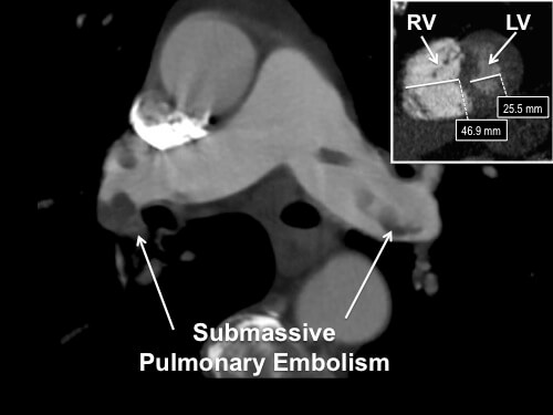
81 year old female with history of recently diagnosed pancreatic/duodenal mass that presented with hypoxia, tachycardia, tachypnea, and chest pain. Multipath Curved Planar Reformat image displays extensive pulmonary emboli. Insert (upper right) shows the increased diameter of the Right Ventricle (RV) vs. the Left Ventricle (LV). Producing these images requires advanced software and skilled technologists available in our lab.

Nancy Ware
3DQ Technologist