 https://3dqlab.stanford.edu/wp-content/uploads/2025/05/diep300.png
300
300
Kyle Gifford
https://3dqlab.stanford.edu/wp-content/uploads/2023/08/3DQ-Website-Logo-Header3.png
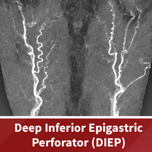
Kyle Gifford2025-05-27 10:36:282025-05-30 10:48:36DIEP (Deep Inferior Epigastric Perforator) Flap Reconstruction
https://3dqlab.stanford.edu/wp-content/uploads/2025/05/diep300.png
300
300
Kyle Gifford
https://3dqlab.stanford.edu/wp-content/uploads/2023/08/3DQ-Website-Logo-Header3.png
Kyle Gifford2025-05-27 10:36:282025-05-30 10:48:36DIEP (Deep Inferior Epigastric Perforator) Flap Reconstruction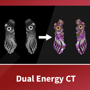 https://3dqlab.stanford.edu/wp-content/uploads/2025/05/dual-energy-ct-300.png
300
300
Kyle Gifford
https://3dqlab.stanford.edu/wp-content/uploads/2023/08/3DQ-Website-Logo-Header3.png
Kyle Gifford2025-05-15 14:05:252025-05-21 10:16:51Dual Energy CT
https://3dqlab.stanford.edu/wp-content/uploads/2025/05/dual-energy-ct-300.png
300
300
Kyle Gifford
https://3dqlab.stanford.edu/wp-content/uploads/2023/08/3DQ-Website-Logo-Header3.png
Kyle Gifford2025-05-15 14:05:252025-05-21 10:16:51Dual Energy CT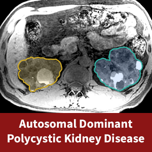 https://3dqlab.stanford.edu/wp-content/uploads/2025/03/adpkd-300.png
300
300
Kyle Gifford
https://3dqlab.stanford.edu/wp-content/uploads/2023/08/3DQ-Website-Logo-Header3.png
Kyle Gifford2025-03-10 15:50:032025-03-13 13:56:53Case of the Month – Autosomal Dominant Polycystic Kidney Disease (ADPKD)
https://3dqlab.stanford.edu/wp-content/uploads/2025/03/adpkd-300.png
300
300
Kyle Gifford
https://3dqlab.stanford.edu/wp-content/uploads/2023/08/3DQ-Website-Logo-Header3.png
Kyle Gifford2025-03-10 15:50:032025-03-13 13:56:53Case of the Month – Autosomal Dominant Polycystic Kidney Disease (ADPKD) https://3dqlab.stanford.edu/wp-content/uploads/2024/12/Prostate-Embolization-300.png
300
300
Kyle Gifford
https://3dqlab.stanford.edu/wp-content/uploads/2023/08/3DQ-Website-Logo-Header3.png
Kyle Gifford2024-12-06 12:39:462024-12-30 10:41:30Case of the Month – Prostate Embolization
https://3dqlab.stanford.edu/wp-content/uploads/2024/12/Prostate-Embolization-300.png
300
300
Kyle Gifford
https://3dqlab.stanford.edu/wp-content/uploads/2023/08/3DQ-Website-Logo-Header3.png
Kyle Gifford2024-12-06 12:39:462024-12-30 10:41:30Case of the Month – Prostate Embolization https://3dqlab.stanford.edu/wp-content/uploads/2024/01/nancy-300.png
300
300
Kyle Gifford
https://3dqlab.stanford.edu/wp-content/uploads/2023/08/3DQ-Website-Logo-Header3.png
Kyle Gifford2024-01-08 15:31:152024-01-11 12:14:35Get to Know the 3D Pro: Nancy Ware
https://3dqlab.stanford.edu/wp-content/uploads/2024/01/nancy-300.png
300
300
Kyle Gifford
https://3dqlab.stanford.edu/wp-content/uploads/2023/08/3DQ-Website-Logo-Header3.png
Kyle Gifford2024-01-08 15:31:152024-01-11 12:14:35Get to Know the 3D Pro: Nancy Ware https://3dqlab.stanford.edu/wp-content/uploads/2023/12/vascular-distance-300.jpg
300
300
Kyle Gifford
https://3dqlab.stanford.edu/wp-content/uploads/2023/08/3DQ-Website-Logo-Header3.png
Kyle Gifford2023-12-15 13:57:402023-12-28 15:19:36Vascular Distance Measurement
https://3dqlab.stanford.edu/wp-content/uploads/2023/12/vascular-distance-300.jpg
300
300
Kyle Gifford
https://3dqlab.stanford.edu/wp-content/uploads/2023/08/3DQ-Website-Logo-Header3.png
Kyle Gifford2023-12-15 13:57:402023-12-28 15:19:36Vascular Distance Measurement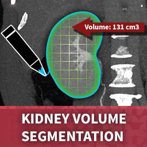 https://3dqlab.stanford.edu/wp-content/uploads/2023/12/kidney-volume-300.jpg
300
300
Kyle Gifford
https://3dqlab.stanford.edu/wp-content/uploads/2023/08/3DQ-Website-Logo-Header3.png
Kyle Gifford2023-12-08 16:00:362024-01-04 15:46:30Kidney Volume
https://3dqlab.stanford.edu/wp-content/uploads/2023/12/kidney-volume-300.jpg
300
300
Kyle Gifford
https://3dqlab.stanford.edu/wp-content/uploads/2023/08/3DQ-Website-Logo-Header3.png
Kyle Gifford2023-12-08 16:00:362024-01-04 15:46:30Kidney Volume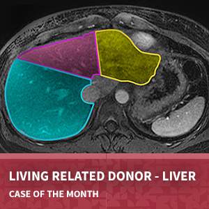 https://3dqlab.stanford.edu/wp-content/uploads/2023/12/LRD-Thumbnail.jpg
300
300
Kyle Gifford
https://3dqlab.stanford.edu/wp-content/uploads/2023/08/3DQ-Website-Logo-Header3.png
Kyle Gifford2023-12-01 12:04:332023-12-07 15:42:27Living Related Donor – Liver
https://3dqlab.stanford.edu/wp-content/uploads/2023/12/LRD-Thumbnail.jpg
300
300
Kyle Gifford
https://3dqlab.stanford.edu/wp-content/uploads/2023/08/3DQ-Website-Logo-Header3.png
Kyle Gifford2023-12-01 12:04:332023-12-07 15:42:27Living Related Donor – Liver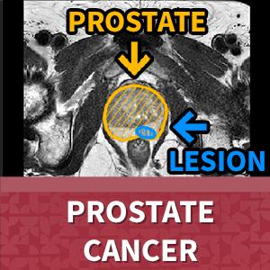 https://3dqlab.stanford.edu/wp-content/uploads/2023/11/prostate-cancer-300.jpg
300
300
Kyle Gifford
https://3dqlab.stanford.edu/wp-content/uploads/2023/08/3DQ-Website-Logo-Header3.png
Kyle Gifford2023-11-08 17:09:162023-11-15 13:56:07Prostate Cancer
https://3dqlab.stanford.edu/wp-content/uploads/2023/11/prostate-cancer-300.jpg
300
300
Kyle Gifford
https://3dqlab.stanford.edu/wp-content/uploads/2023/08/3DQ-Website-Logo-Header3.png
Kyle Gifford2023-11-08 17:09:162023-11-15 13:56:07Prostate Cancer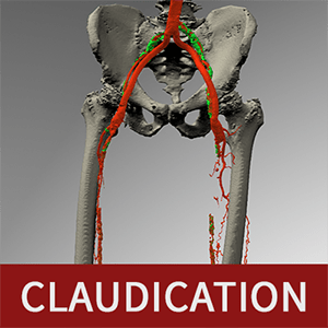 https://3dqlab.stanford.edu/wp-content/uploads/2023/06/claudicationtitlesmall.png
300
300
Kyle Gifford
https://3dqlab.stanford.edu/wp-content/uploads/2023/08/3DQ-Website-Logo-Header3.png
Kyle Gifford2023-06-02 16:15:362023-09-17 19:48:11Claudication
https://3dqlab.stanford.edu/wp-content/uploads/2023/06/claudicationtitlesmall.png
300
300
Kyle Gifford
https://3dqlab.stanford.edu/wp-content/uploads/2023/08/3DQ-Website-Logo-Header3.png
Kyle Gifford2023-06-02 16:15:362023-09-17 19:48:11Claudication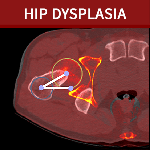 https://3dqlab.stanford.edu/wp-content/uploads/2023/05/title6.png
300
300
Kyle Gifford
https://3dqlab.stanford.edu/wp-content/uploads/2023/08/3DQ-Website-Logo-Header3.png
Kyle Gifford2023-05-03 14:37:302023-09-18 09:01:26Hip Preservation
https://3dqlab.stanford.edu/wp-content/uploads/2023/05/title6.png
300
300
Kyle Gifford
https://3dqlab.stanford.edu/wp-content/uploads/2023/08/3DQ-Website-Logo-Header3.png
Kyle Gifford2023-05-03 14:37:302023-09-18 09:01:26Hip Preservation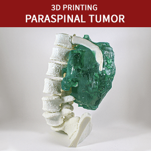 https://3dqlab.stanford.edu/wp-content/uploads/2023/03/0323-cotm-feature.png
300
300
Kyle Gifford
https://3dqlab.stanford.edu/wp-content/uploads/2023/08/3DQ-Website-Logo-Header3.png
Kyle Gifford2023-03-29 09:38:022023-09-19 08:18:253D Printing – Paraspinal Tumor
https://3dqlab.stanford.edu/wp-content/uploads/2023/03/0323-cotm-feature.png
300
300
Kyle Gifford
https://3dqlab.stanford.edu/wp-content/uploads/2023/08/3DQ-Website-Logo-Header3.png
Kyle Gifford2023-03-29 09:38:022023-09-19 08:18:253D Printing – Paraspinal Tumor https://3dqlab.stanford.edu/wp-content/uploads/2019/06/Breast-Recon.jpeg
836
1000
shanwalt
https://3dqlab.stanford.edu/wp-content/uploads/2023/08/3DQ-Website-Logo-Header3.png
shanwalt2019-06-21 14:46:222023-09-19 08:21:09April 2019: 3D Print for Breast Reconstruction Planning
https://3dqlab.stanford.edu/wp-content/uploads/2019/06/Breast-Recon.jpeg
836
1000
shanwalt
https://3dqlab.stanford.edu/wp-content/uploads/2023/08/3DQ-Website-Logo-Header3.png
shanwalt2019-06-21 14:46:222023-09-19 08:21:09April 2019: 3D Print for Breast Reconstruction Planning https://3dqlab.stanford.edu/wp-content/uploads/2023/08/3DQ-Website-Logo-Header3.png
0
0
cletrong
https://3dqlab.stanford.edu/wp-content/uploads/2023/08/3DQ-Website-Logo-Header3.png
cletrong2018-06-07 09:17:212023-09-19 08:27:03Cholelithiasis (Gallstones)
https://3dqlab.stanford.edu/wp-content/uploads/2023/08/3DQ-Website-Logo-Header3.png
0
0
cletrong
https://3dqlab.stanford.edu/wp-content/uploads/2023/08/3DQ-Website-Logo-Header3.png
cletrong2018-06-07 09:17:212023-09-19 08:27:03Cholelithiasis (Gallstones) https://3dqlab.stanford.edu/wp-content/uploads/2017/09/all-1.png
573
800
cletrong
https://3dqlab.stanford.edu/wp-content/uploads/2023/08/3DQ-Website-Logo-Header3.png
cletrong2017-09-26 12:32:312023-09-19 08:34:33Virtual Colonoscopy
https://3dqlab.stanford.edu/wp-content/uploads/2017/09/all-1.png
573
800
cletrong
https://3dqlab.stanford.edu/wp-content/uploads/2023/08/3DQ-Website-Logo-Header3.png
cletrong2017-09-26 12:32:312023-09-19 08:34:33Virtual Colonoscopy https://3dqlab.stanford.edu/wp-content/uploads/2017/08/fused.gif
800
800
cletrong
https://3dqlab.stanford.edu/wp-content/uploads/2023/08/3DQ-Website-Logo-Header3.png
cletrong2017-08-14 14:54:102023-09-19 08:33:06August 2017 – Cross-Fused Renal Ectopia
https://3dqlab.stanford.edu/wp-content/uploads/2017/08/fused.gif
800
800
cletrong
https://3dqlab.stanford.edu/wp-content/uploads/2023/08/3DQ-Website-Logo-Header3.png
cletrong2017-08-14 14:54:102023-09-19 08:33:06August 2017 – Cross-Fused Renal Ectopia https://3dqlab.stanford.edu/wp-content/uploads/2016/11/Capture3.png
612
821
cletrong
https://3dqlab.stanford.edu/wp-content/uploads/2023/08/3DQ-Website-Logo-Header3.png
cletrong2016-11-10 12:33:082023-09-19 08:39:13November 2016 – Living Related Renal Donor
https://3dqlab.stanford.edu/wp-content/uploads/2016/11/Capture3.png
612
821
cletrong
https://3dqlab.stanford.edu/wp-content/uploads/2023/08/3DQ-Website-Logo-Header3.png
cletrong2016-11-10 12:33:082023-09-19 08:39:13November 2016 – Living Related Renal Donor https://3dqlab.stanford.edu/wp-content/uploads/2016/06/pre-1.jpeg
400
400
cletrong
https://3dqlab.stanford.edu/wp-content/uploads/2023/08/3DQ-Website-Logo-Header3.png
cletrong2016-06-16 10:59:252023-09-19 08:42:27June 2016
https://3dqlab.stanford.edu/wp-content/uploads/2016/06/pre-1.jpeg
400
400
cletrong
https://3dqlab.stanford.edu/wp-content/uploads/2023/08/3DQ-Website-Logo-Header3.png
cletrong2016-06-16 10:59:252023-09-19 08:42:27June 2016 https://3dqlab.stanford.edu/wp-content/uploads/2016/05/Normal-Renal-Artery_1-768x768-1.jpeg
768
768
cletrong
https://3dqlab.stanford.edu/wp-content/uploads/2023/08/3DQ-Website-Logo-Header3.png
cletrong2016-05-17 07:50:302023-09-19 08:42:15May 2016
https://3dqlab.stanford.edu/wp-content/uploads/2023/08/3DQ-Website-Logo-Header3.png
0
0
cletrong
https://3dqlab.stanford.edu/wp-content/uploads/2023/08/3DQ-Website-Logo-Header3.png
cletrong2016-02-16 11:33:102023-09-19 08:44:47January 2016
https://3dqlab.stanford.edu/wp-content/uploads/2016/05/Normal-Renal-Artery_1-768x768-1.jpeg
768
768
cletrong
https://3dqlab.stanford.edu/wp-content/uploads/2023/08/3DQ-Website-Logo-Header3.png
cletrong2016-05-17 07:50:302023-09-19 08:42:15May 2016
https://3dqlab.stanford.edu/wp-content/uploads/2023/08/3DQ-Website-Logo-Header3.png
0
0
cletrong
https://3dqlab.stanford.edu/wp-content/uploads/2023/08/3DQ-Website-Logo-Header3.png
cletrong2016-02-16 11:33:102023-09-19 08:44:47January 2016 https://3dqlab.stanford.edu/wp-content/uploads/2015/11/Normal-clipped-1.jpeg
316
600
cletrong
https://3dqlab.stanford.edu/wp-content/uploads/2023/08/3DQ-Website-Logo-Header3.png
cletrong2015-11-10 11:16:422023-09-19 08:47:50September 2015
https://3dqlab.stanford.edu/wp-content/uploads/2015/11/Normal-clipped-1.jpeg
316
600
cletrong
https://3dqlab.stanford.edu/wp-content/uploads/2023/08/3DQ-Website-Logo-Header3.png
cletrong2015-11-10 11:16:422023-09-19 08:47:50September 2015 https://3dqlab.stanford.edu/wp-content/uploads/2015/07/Capture-5-1-1.png
600
600
cletrong
https://3dqlab.stanford.edu/wp-content/uploads/2023/08/3DQ-Website-Logo-Header3.png
cletrong2015-07-24 11:04:072023-09-19 08:49:34June 2015
https://3dqlab.stanford.edu/wp-content/uploads/2015/07/Capture-5-1-1.png
600
600
cletrong
https://3dqlab.stanford.edu/wp-content/uploads/2023/08/3DQ-Website-Logo-Header3.png
cletrong2015-07-24 11:04:072023-09-19 08:49:34June 2015 https://3dqlab.stanford.edu/wp-content/uploads/2015/01/1501COTM-Linda1-1.png
450
600
admin
https://3dqlab.stanford.edu/wp-content/uploads/2023/08/3DQ-Website-Logo-Header3.png
admin2015-01-27 22:28:302023-09-19 08:54:47January 2015
https://3dqlab.stanford.edu/wp-content/uploads/2015/01/1501COTM-Linda1-1.png
450
600
admin
https://3dqlab.stanford.edu/wp-content/uploads/2023/08/3DQ-Website-Logo-Header3.png
admin2015-01-27 22:28:302023-09-19 08:54:47January 2015 https://3dqlab.stanford.edu/wp-content/uploads/2014/08/Slide1-copy-2-1.png
375
500
admin
https://3dqlab.stanford.edu/wp-content/uploads/2023/08/3DQ-Website-Logo-Header3.png
admin2014-08-29 19:13:102023-09-19 08:57:20August 2014
https://3dqlab.stanford.edu/wp-content/uploads/2014/08/Slide1-copy-2-1.png
375
500
admin
https://3dqlab.stanford.edu/wp-content/uploads/2023/08/3DQ-Website-Logo-Header3.png
admin2014-08-29 19:13:102023-09-19 08:57:20August 2014 https://3dqlab.stanford.edu/wp-content/uploads/2014/07/Slide11-1.png
375
500
admin
https://3dqlab.stanford.edu/wp-content/uploads/2023/08/3DQ-Website-Logo-Header3.png
admin2014-07-01 19:18:442023-09-19 08:59:11June 2014
https://3dqlab.stanford.edu/wp-content/uploads/2014/07/Slide11-1.png
375
500
admin
https://3dqlab.stanford.edu/wp-content/uploads/2023/08/3DQ-Website-Logo-Header3.png
admin2014-07-01 19:18:442023-09-19 08:59:11June 2014 https://3dqlab.stanford.edu/wp-content/uploads/2014/06/Slide12-1.png
375
500
admin
https://3dqlab.stanford.edu/wp-content/uploads/2023/08/3DQ-Website-Logo-Header3.png
admin2014-06-03 19:20:272023-09-19 08:59:20May 2014
https://3dqlab.stanford.edu/wp-content/uploads/2014/06/Slide12-1.png
375
500
admin
https://3dqlab.stanford.edu/wp-content/uploads/2023/08/3DQ-Website-Logo-Header3.png
admin2014-06-03 19:20:272023-09-19 08:59:20May 2014 https://3dqlab.stanford.edu/wp-content/uploads/2013/11/Slide15-1.png
375
500
admin
https://3dqlab.stanford.edu/wp-content/uploads/2023/08/3DQ-Website-Logo-Header3.png
admin2013-11-26 19:28:192023-09-19 09:00:37December 2013
https://3dqlab.stanford.edu/wp-content/uploads/2013/11/Slide15-1.png
375
500
admin
https://3dqlab.stanford.edu/wp-content/uploads/2023/08/3DQ-Website-Logo-Header3.png
admin2013-11-26 19:28:192023-09-19 09:00:37December 2013 https://3dqlab.stanford.edu/wp-content/uploads/2013/11/Nov_sliec500x375-1.png
375
500
admin
https://3dqlab.stanford.edu/wp-content/uploads/2023/08/3DQ-Website-Logo-Header3.png
admin2013-11-04 19:30:222023-09-19 09:01:03November 2013
https://3dqlab.stanford.edu/wp-content/uploads/2013/04/Kesh-March2013-1.png
375
500
admin
https://3dqlab.stanford.edu/wp-content/uploads/2023/08/3DQ-Website-Logo-Header3.png
admin2013-04-26 19:44:192023-10-11 15:53:19March 2013
https://3dqlab.stanford.edu/wp-content/uploads/2013/11/Nov_sliec500x375-1.png
375
500
admin
https://3dqlab.stanford.edu/wp-content/uploads/2023/08/3DQ-Website-Logo-Header3.png
admin2013-11-04 19:30:222023-09-19 09:01:03November 2013
https://3dqlab.stanford.edu/wp-content/uploads/2013/04/Kesh-March2013-1.png
375
500
admin
https://3dqlab.stanford.edu/wp-content/uploads/2023/08/3DQ-Website-Logo-Header3.png
admin2013-04-26 19:44:192023-10-11 15:53:19March 2013