
3D Printing – Paraspinal Tumor
A patient with a large tumor near the spine, ribs, and hip region was brought to the attention of the 3DQ Lab by the surgical oncology team. The tumor was complex and beginning to invade the bone and spinal cord. This complexity prompted the surgeon to explore advanced visualization technologies, thereby requesting a 3D print. These tumors are known as paraspinal tumors, which are soft tissue growths that may invade the vertebral column, major nerve roots, and blood vessels. The impact of the tumor growth may cause symptoms such as pain, weakness, and numbness in the back, arms, and legs. Treatment for this case was to remove the tumor in surgery and apply radiation therapy afterward to destroy possible remaining cancer cells.
Figure A (Right): This rotational 3D visualization represents the information that was 3D-printed. The spine (L5 through T12 vertebrae), 12th rib, and the superior portion of the pelvis are displayed in grey and the paraspinal tumor is displayed in translucent green. The bubble-like structures inside the translucent green area are calcifications in the tumor.

CT and MRI scans were acquired for this patient since CTs capture bone details clearly and MRI is ideal for soft tissue like the tumor. The anatomy to be included in the model was extracted (segmented) by a trained radiologic technologist, then verified by an oncologist for accuracy. The approved segmentation was then extracted to a data format used in 3D printing known as STL.
Learn more about segmentation here: https://3dqlab.stanford.edu/technique-of-the-week-segmentation/

Figure B: Segmentation of the bony anatomy (yellow) was performed on the CT.

Figure C: Segmentation of the tumor (green) was performed on the MRI.
The 3D-printed model was created using polyjet technology since the complexity of the anatomic relationships benefit from multi-color and transparent materials. Surgeons reviewed the 3D print, marking potential cut planes for resection. The 3D print helped the surgical team validate the treatment plan by offering additional perspectives prior to the surgical procedure.

Figure D: The completed 3D print.
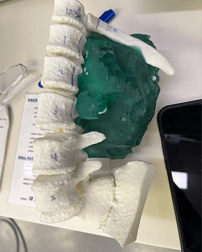
Figure E: The 3D print, seen with marked vertebrae and lines showing potential resection boundaries.

