
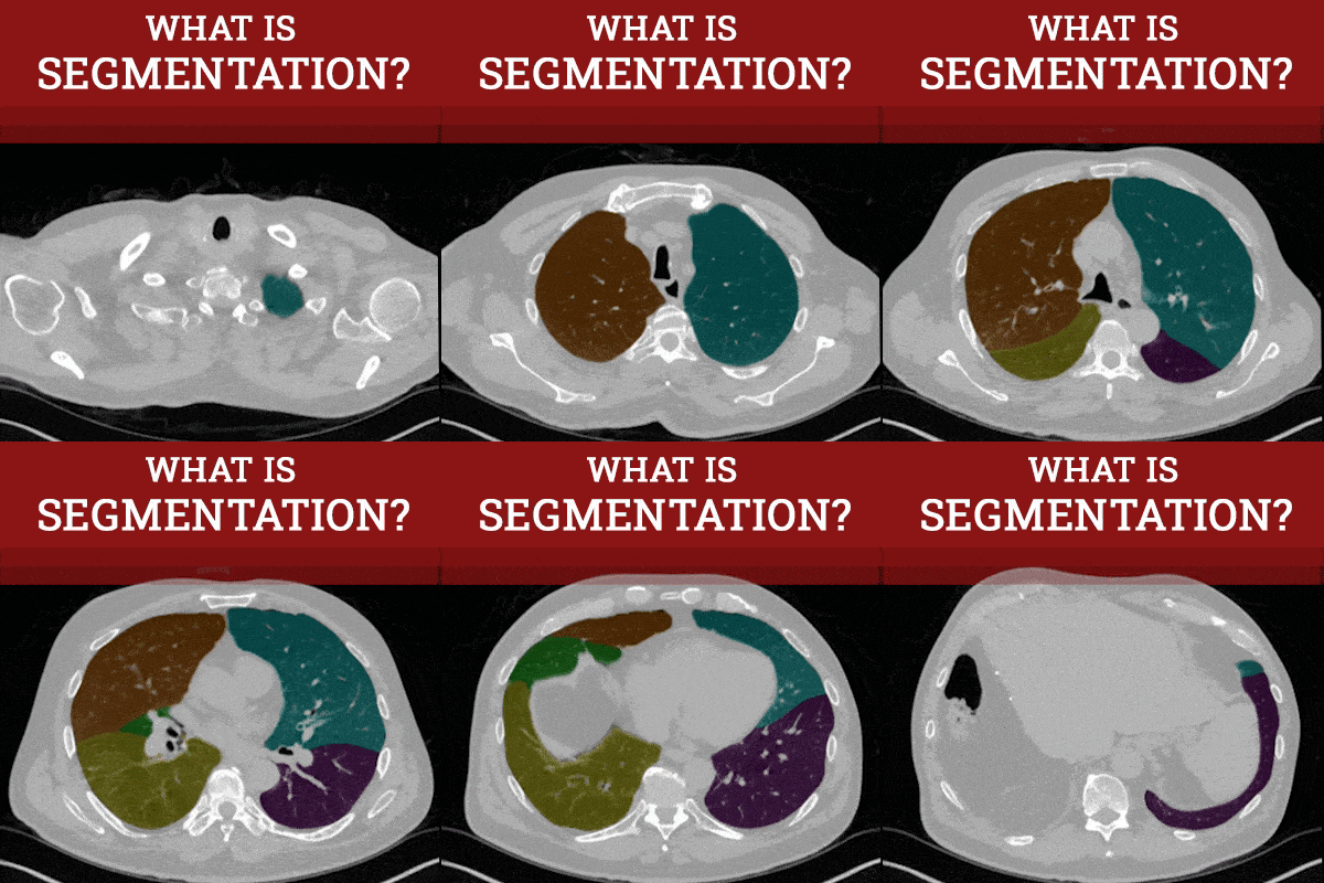

Medical image segmentation is a process that involves separating patient scan data images into regions based on different properties. These regions can represent different anatomical structures or tissues, such as bones, organs, implanted hardware, or tumors. By accurately identifying these regions, they can be helpful for diagnosis, treatment planning, and a better understanding of an individual’s unique anatomy.

Segmentation is often performed by computer algorithms that are guided by certified human technologists. There are many different approaches, both automatic and manual, but some common ones include:
Thresholding: Thresholding selects portions of patient scan data by setting a value range based on pixel intensity (Hounsfield units). This is useful for selecting large regions of similar pixel intensity, such as bone, air or contrast-enhanced blood flow.
Figure A (Right) An axial MRI demonstrating the range of thresholding across cranial anatomy.

Region Growing: Region growing involves selecting a seed point in the patient data, and expanding that selection outward to nearby pixels with similar Hounsfield units. This technique is useful for segmenting winding anatomy with relatively similar pixel intensity, such as blood vessels.
Figure B (Right) Region growing is being performed on this sagittal vessel by selecting the bright contrast of the vessel as the seed point and expanding the selection to nearby similar densities, thus following the path of the vessel.

Manual Segmentation: Manual segmentation is a technique where a human expert manually marks the regions of interest in patient scan data. This usually involves drawing boundaries slice by slice with brush and lasso tools. This process is often time consuming, labor-intensive, and can result in some variability depending on the user’s expertise.
Figure C (Right) This example demonstrates a vessel with uneven contrast being selected by hand with a lasso tool.

Machine Learning: This technique involves training a machine learning algorithm to segment image data based on certain preset characteristics.
Figure D (Below) The left clip demonstrates emphysema that has been segmented automatically in red with AI. The right image is a report that is automatically generated from the segmented data, demonstrating the spread of the disease.


The final result of segmentation demonstrates an isolated portion of patient anatomy which is used for diagnosis and treatment planning, research, education, and visualization.


Figure E (Above): The left two videos demonstrate the final segmentation and rotational videos of a living-related donor kidney for transplant purposes. The right two images show a segmented pelvis ilium, which is useful for preop planning for fixation of fractures, and postoperative assesment of hardware or bony growth.

