
The TMVR evaluation begins with a 3DQ Lab technologist obtaining the imaging study for this patient, which is a cardiac-gated CT Angiogram (CTA) of the chest, abdomen, and pelvis, with contrast to visualize blood flow. Cardiac gating synchronizes the image acquisition with specific phases of the heart’s cycle, allowing for a clearer and more detailed view of the heart’s structure and function.

Figure B: A 4D, four-perspective view of the mitral valve offers a comprehensive view of the valve in motion throughout the cardiac cycle. This is useful for evaluating valve morphology and function.
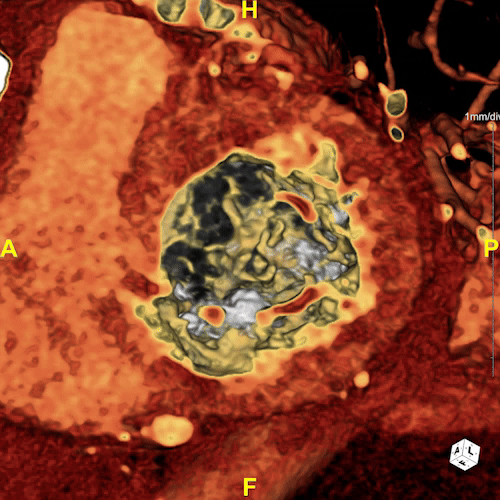
Figure C: This 4D video captures the valve’s dynamics throughout the cardiac cycle, displaying its opening and closing movements. This visualization can be used for detecting calcium buildup and other irregularities such as regurgitation.
Our Lab provided the below imaging and measurements to the intervention team; the extensive calcifications lead to the evaluation of two different valve devices. Annulus measurements were made to capture the dimensions of the mitral valve’s opening during different cardiac phases, which is important for selecting a prosthetic valve that matches the patient’s anatomical requirements. By measuring the annulus perimeter, area, and diameters, we ensure that the chosen valve not only fits securely but also functions optimally within the structural constraints of each patient’s heart. Additionally, these measurements allow virtual placement of the valve, allowing us to simulate various positioning scenarios to predict and optimize how the valve will function post-implantation.
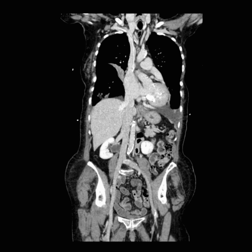
Figure D: A CPR (Curved Planar Reformation) of the right common femoral vein through the superior vena cava presents a flattened view of the venous delivery pathway in a single image. This method is useful for assessing the veins to ensure safe catheter access, displaying the path that the catheter will take during the TMVR procedure.
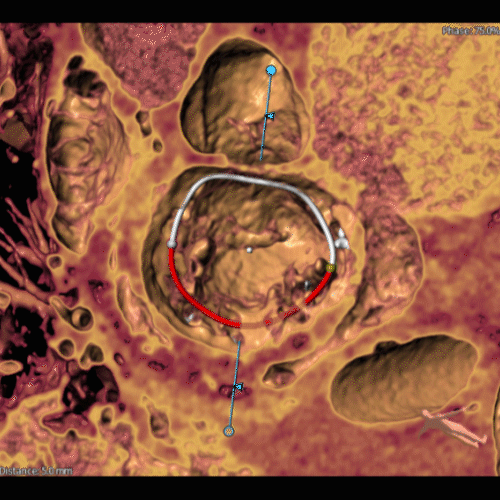
Figure E: Measurements specific to the Intrepid valve, which is tailor-made for mitral valve replacement, were provided to the TMVR team. These measurements are useful in selecting the correct size and type of valve for the patient, ensuring that it fits properly and functions effectively after it is deployed.

Figure F: Measurements for the Sapien valve, originally developed for transcatheter aortic valve replacements, were also provided. The Sapien valve offers a less invasive alternative for patients who have undergone previous valve surgeries or who are at high risk for open-heart surgery.

