3D and Quantitative Imaging Laboratory
  • Home
  • Education
    • Education
    • Educational Post Feed
  • Research
    • Research
    • Validation Cohort: Acute Uncomplicated Type-B Aortic Dissection
  • Patient Care
    • Patient Care
    • 3D Printing in Medicine
    • 3DQ Lab TRAC Team (Tumor Response)
  • Case Studies
    • Case Studies
    • Innovation Projects
  • Click to open the search input field Click to open the search input field Search
  • Menu Menu

Lumbar Puncture Training Models

Collaborators: Stanford General Surgery

Lumbar puncture training often relies on physical phantom models that allow learners to practice needle placement before performing procedures on patients. Commercial training models can be costly and may offer limited opportunities for customization.

To explore an alternative approach, the Stanford 3DQ Lab collaborated with Radiology residents and Stanford General Surgery to develop a reusable lumbar puncture phantom for fluoroscopy-guided training. The project focused on creating a lower-cost model that could be manufactured in-house while maintaining realistic imaging characteristics and procedural functionality.

Figure A: 3D render of the provided 3D printed training models to be casted in ballistics gel.
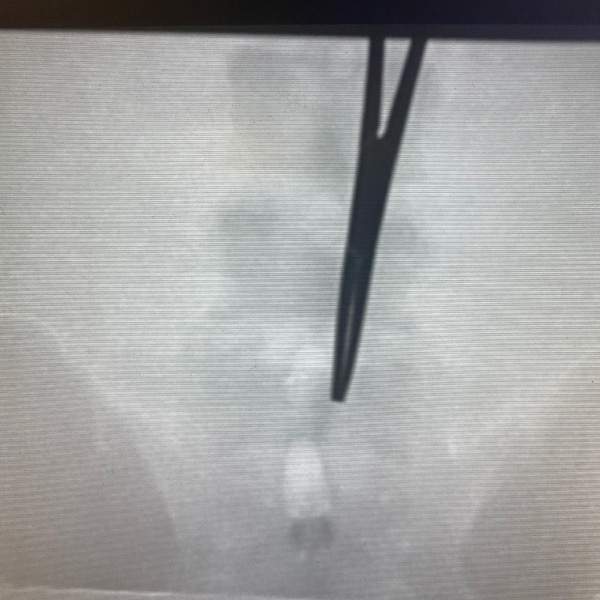
Figure B. Fluoroscopic image of the lumbar puncture training phantom during simulated needle placement.

Development involved evaluating multiple 3D printing materials and construction techniques to identify a suitable balance between durability, radiographic appearance, and procedural realism. The team worked closely with material manufacturers and end users throughout the design process. ABS was ultimately selected for anatomical components, and different infill patterns and densities were explored to better replicate vertebral anatomy under fluoroscopy. A fluid-filled spinal canal and cast materials were incorporated to support repeated training use.

The resulting phantom provided a customizable platform for fluoroscopy-guided lumbar puncture education while demonstrating how 3D printing could be used to create institution-specific training tools.

Categories

  • Head & Neck
  • Chest
  • Abdomen & Pelvis
  • Upper Extremity
  • Lower Extremity
  • Case of the Month
  • Techniques

3DQ Lab – Grant Building

300 Pasteur Drive
Stanford, CA 94305

(650) 725-8432

Directions to Grant

3DQ Lab – Clark Building

318 Campus Drive
Stanford, CA 94305

(650) 725-6862

Directions to Clark

Learn More About the Lab

  • About Us
  • Meet the Team
Search Search

Copyright © Stanford University

© Copyright - 3D and Quantitative Imaging Laboratory - Enfold Theme by Kriesi
  • Link to LinkedIn
  • Link to Instagram
Link to: DICOM CVS Reports Link to: DICOM CVS Reports DICOM CVS Reports Link to: AI Evaluation and Deployment Link to: AI Evaluation and Deployment AI Evaluation and Deployment
Scroll to top Scroll to top Scroll to top