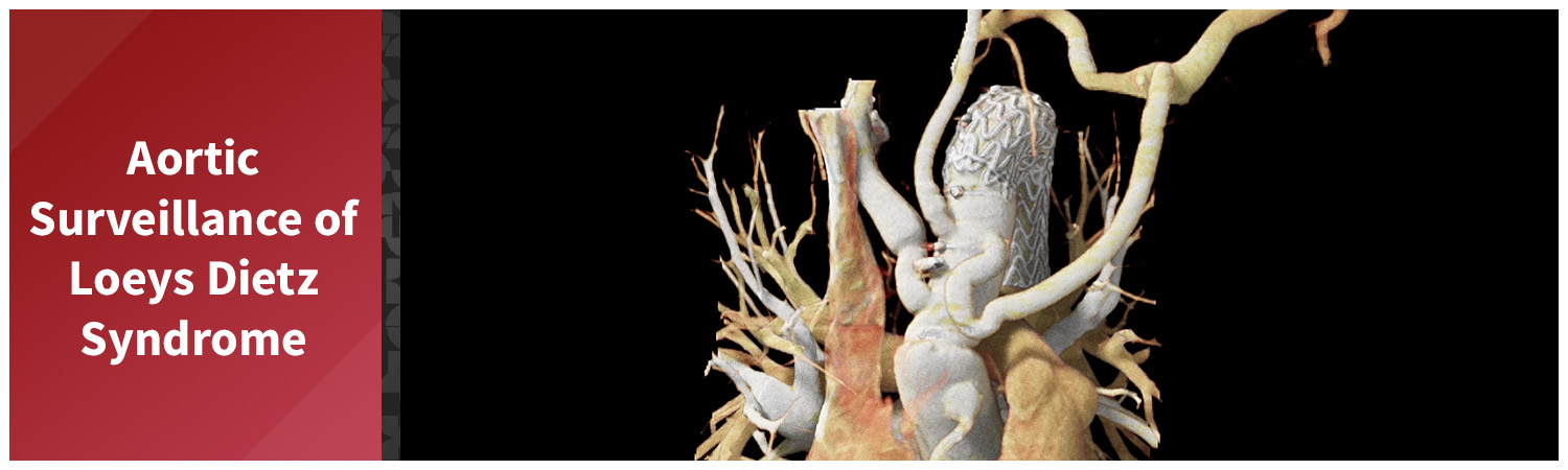
A patient with a connective tissue disorder was referred to the 3DQ Lab for longitudinal aortic surveillance. In conditions such as Loeys-Dietz syndrome, the wall of the aorta can be weaker than normal, allowing portions of the vessel to enlarge over time. Because progressive enlargement can increase the risk of dissection or rupture, serial imaging and standardized measurements are used to monitor changes and help guide treatment decisions.
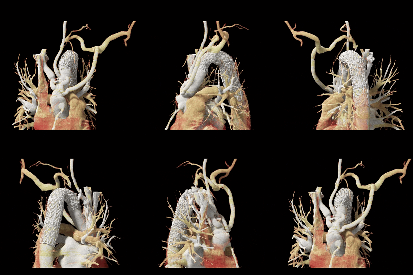
This case was particularly challenging because the patient had undergone multiple prior aortic repairs. Portions of the aorta had been replaced with surgical graft material or treated with an endovascular stent, and some vascular reconstructions had been performed to maintain blood flow to critical arteries. While these repairs can be lifesaving, they significantly alter the anatomy and can make it difficult to obtain reproducible measurements from one examination to the next.
Figure A (Right): Rotational volume render of the aortic arch demonstrating prior surgical repair, endovascular stenting, and bypass graft reconstruction.
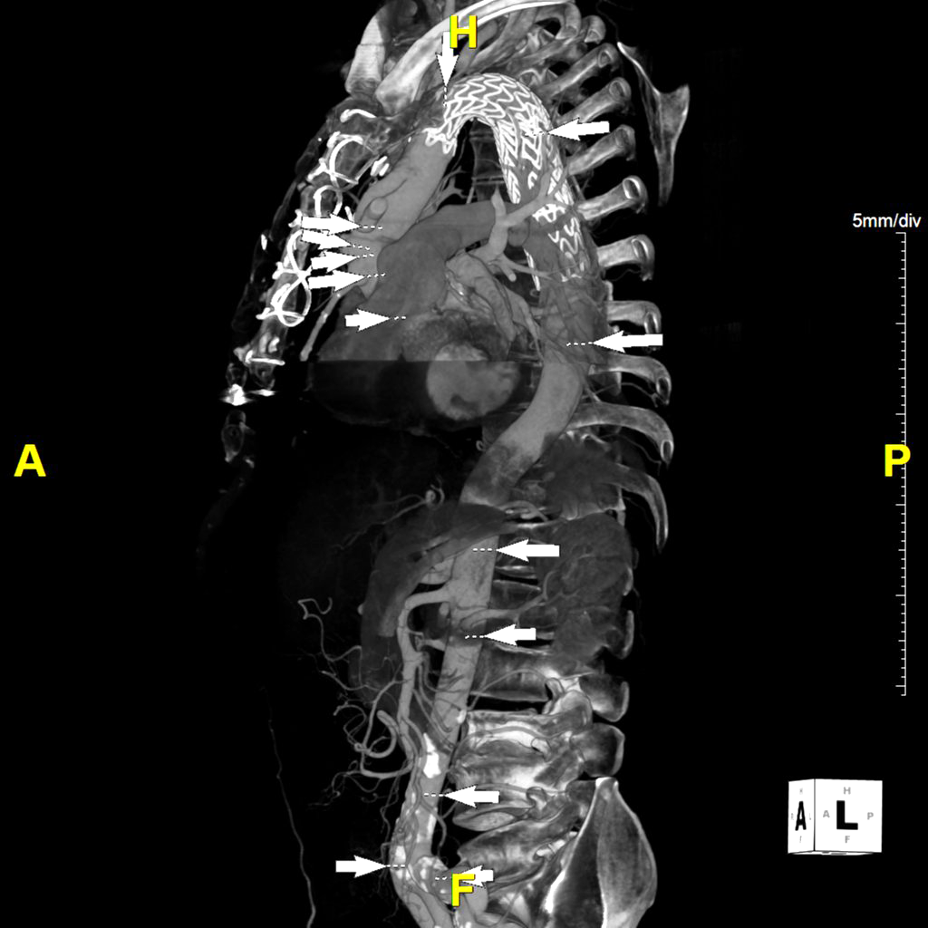
To support accurate longitudinal follow-up, the 3DQ Lab performs measurements at standardized locations during every examination. These exact measurement locations are used across all of the patient’s studies, allowing clinicians to accurately compare changes over time. In repaired segments, measurements are obtained based on the anatomy as it exists today. When thrombus is present, both the lumen and thrombus can be evaluated to provide a more complete assessment of change over time.
Learn more about longitudinal analysis here.
Figure B (Left): White arrows indicating standardized measurement locations used during longitudinal aortic surveillance. Measurements are obtained at the same anatomical stations during each visit to support consistent comparison over time.
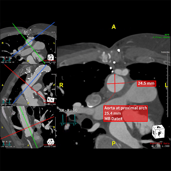
Figure C: Cross-sectional measurement obtained at a standardized surveillance location within the stented segment of the aorta.
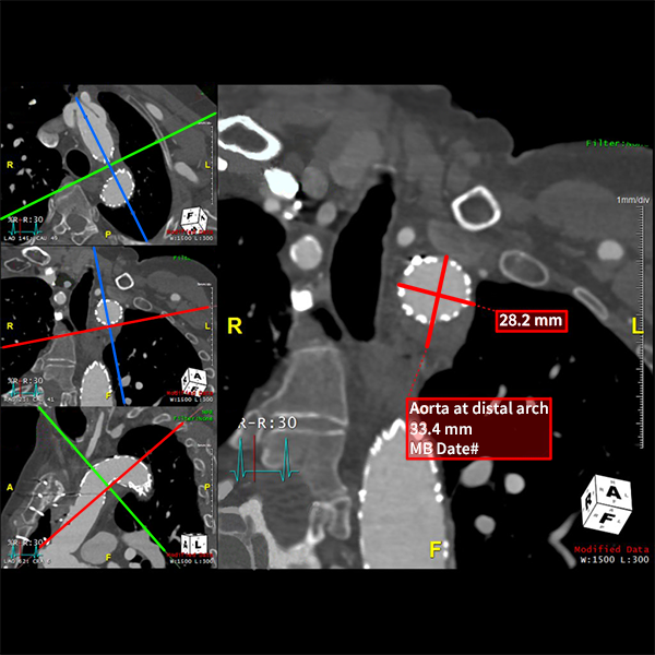
Figure D: Cross-sectional measurement obtained at a standardized surveillance location adjacent to the bypass graft reconstruction.
Complex repairs can also affect how measurement planes are created. Grafts, stents, and postoperative changes may make it difficult to achieve a perfectly orthogonal measurement plane, requiring careful adjustment while still maintaining the target anatomical location. When a measurement cannot be obtained reliably because of postoperative anatomy or imaging artifacts, the limitation is documented and communicated to the clinical team to provide appropriate context for interpretation.
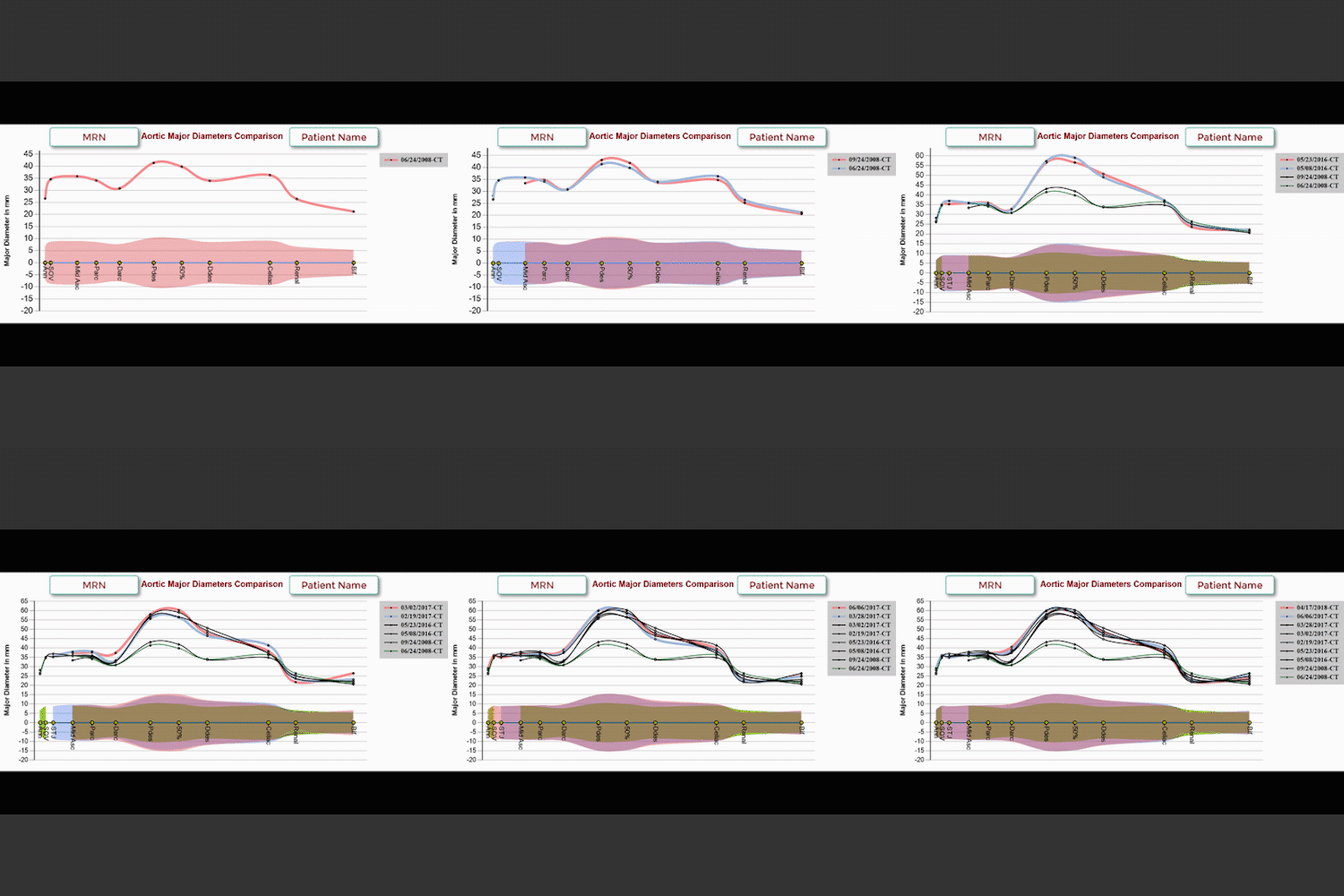
All measurements were incorporated into a longitudinal diameter graph showing how each segment of the aorta has changed over time. Consolidating measurements from individual imaging studies into a single graph allows clinicians to more easily identify which regions are stable and which may be growing.
Figure E: Example graph of the standardized aortic measurements, visualizing changes of the aorta over time.