3D and Quantitative Imaging Laboratory
  • Home
  • Education
    • Education
    • Educational Post Feed
  • Research
    • Research
    • Validation Cohort: Acute Uncomplicated Type-B Aortic Dissection
  • Patient Care
    • Patient Care
    • 3D Printing in Medicine
    • 3DQ Lab TRAC Team (Tumor Response)
  • Case Studies
    • Case Studies
    • Innovation Projects
  • Click to open the search input field Click to open the search input field Search
  • Menu Menu

July 2013

Case of the Month, Chest

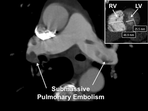
81 year old female with history of recently diagnosed pancreatic/duodenal mass that presented with hypoxia, tachycardia, tachypnea, and chest pain. Multipath Curved Planar Reformat image displays extensive pulmonary emboli. Insert (upper right) shows the increased diameter of the Right Ventricle (RV) vs. the Left Ventricle (LV). Producing these images requires advanced software and skilled technologists available in our lab.

Nancy Ware
3DQ Technologist

July 1, 2013/by admin
https://3dqlab.stanford.edu/wp-content/uploads/2013/07/Caseofthemonth.July2013-1.jpg 375 500 admin https://3dqlab.stanford.edu/wp-content/uploads/2023/08/3DQ-Website-Logo-Header3.png admin2013-07-01 19:39:252023-09-19 09:02:48July 2013

Categories

  • Head & Neck
  • Chest
  • Abdomen & Pelvis
  • Upper Extremity
  • Lower Extremity
  • Case of the Month
  • Techniques

3DQ Lab – Grant Building

300 Pasteur Drive
Stanford, CA 94305

(650) 725-8432

Directions to Grant

3DQ Lab – Clark Building

318 Campus Drive
Stanford, CA 94305

(650) 725-6862

Directions to Clark

Learn More About the Lab

  • About Us
  • Meet the Team
Search Search

Copyright © Stanford University

© Copyright - 3D and Quantitative Imaging Laboratory - Enfold Theme by Kriesi
  • Link to LinkedIn
  • Link to Instagram
Link to: April 2013 Link to: April 2013 April 2013 Link to: August 2013 Link to: August 2013 August 2013
Scroll to top Scroll to top Scroll to top