

While there are various categories of congenital heart defects, each is unique and demands highly specific interventions. This study shows a patient with multiple congenital heart defects who was referred to Radiology and the 3DQ Lab for specialized imaging and visualization. Given the intricacy of the condition, a 3D print was ordered to serve as a visual aid for planning surgical interventions, and as an educational tool for training healthcare professionals who are new to such complex cases.
The patient received a contrast-enhanced CT scan to obtain precise cross-sectional images of their cardiac system. An experienced technologist in the 3DQ Lab meticulously segmented the heart’s various structures, collaborating with a radiologist to pinpoint the specific locations of the cardiac anomalies. The defects that were identified in this unique case are detailed below.
Figure A (Right): Images from an axial CT scan of the heart.
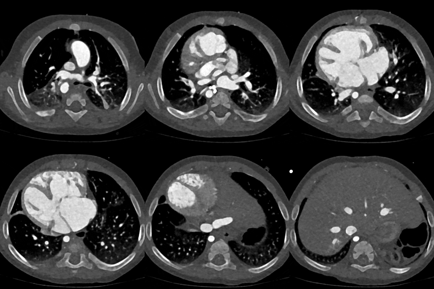
Figure B: 3D reconstruction and animation of the axial CT scan of the heart, with defects shown in color. Teal: Inferior Vena Cava (IVC) Bicaval Connection; Red: Aortic Connection; Magenta: Common Atrioventricular Valve (Approximation); Yellow: Ventricular Septal Defect; Green: Atrial Septal Defect.

Figure C: Primum Atrial Septal Defect (ASD) (Green)
A primum ASD is a type of hole in the wall that separates the upper chambers of the heart (the left and right atria). This hole allows blood to flow between the atria, potentially leading to a mixing of oxygenated and deoxygenated blood.
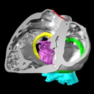
Figure D: Inlet Ventricular Septal Defect (VSD) (Yellow)
An inlet VSD is a hole that occurs in the septum that separates the lower chambers of the heart: the left and right ventricles. This defect allows blood to flow between the ventricles, which can also result in the mixing of oxygenated and deoxygenated blood.
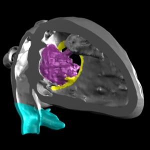
Figure E: Common Atrioventricular Valve (Magenta)
A common atrioventricular valve is a heart defect where the septum that normally separates the left and right atrioventricular valves doesn’t fully form during fetal development. This results in a single shared valve between the upper (atria) and lower (ventricles) heart chambers, instead of the usual two separate valves (mitral on the left and tricuspid on the right).

Figure F: IVC Bicaval Connection (Teal)
This congenital heart condition is characterized by an abnormal connection of the IVC to both atria, rather than its normal connection to the right atrium. It can result in mixing of oxygenated and deoxygenated blood within the heart and is often associated with other cardiac defects.


Figure G: Photos of the completed 3D print.
The segmented data were converted into a 3D mesh file and partitioned into three distinct sections. This division plan was carefully crafted to make sure the central section included the entire conjoined valve structure. The three sections were printed using polyjet technology, a printing method that uses ultraviolet activated liquid polymers to create high resolution and multi-material 3D prints. The conjoined valve was fabricated using a semi-flexible material, while the remaining portions of the model were produced using a rigid, frosty-clear material. After nearly 18 hours of printing, the model was cleaned and delivered to the heart team at Stanford Medicine Children’s Health, who used it to understand and guide the surgical intervention.

