3D and Quantitative Imaging Laboratory
  • Home
  • Education
    • Education
    • Educational Post Feed
  • Research
    • Research
    • Validation Cohort: Acute Uncomplicated Type-B Aortic Dissection
  • Patient Care
    • Patient Care
    • 3D Printing in Medicine
    • 3DQ Lab TRAC Team (Tumor Response)
  • Case Studies
    • Case Studies
  • Search
  • Menu Menu

A young patient arrived at the emergency department after sustaining significant trauma to their lower extremities, resulting in multiple fractures. They had severely comminuted fractures (where the bone is broken into three or more fragments) of the left femur, tibia, and fibula, as well as a comminuted fracture of the right iliac wing, which extended into the sacroiliac joint.

While the fractures were important to assess, the 3DQ Lab’s main concern was visualizing the surrounding vasculature near the fractures to identify potential internal bleeding. The trauma also caused extensive soft tissue injuries, including hematomas beneath the skin and within the muscle, complicating the case.

Figure A (Right): 3D reconstruction of the lower extremities, showing the location of each fracture. This model can be rotated freely by holding and dragging left-click. Clicking on an annotation provides a brief description of the fracture.

The imaging process began with a CT angiogram of the abdomen and pelvis, providing a view of both the bone and vascular anatomy. Using 3D post-processing techniques, the 3DQ Lab generated rotational views of the pelvis, left femur, tibia, and fibula, clearly illustrating the complex fracture patterns (as shown below).

Additionally, a Maximum Intensity Projection (MIP) of the lower extremities was performed to assess blood flow through the arteries and detect any narrowing or vascular obstructions. The 3D Lab employed extensive segmentation efforts to enable clear MIP visualization without interference from bones or other dense structures. This was a significant technical challenge due to the proximity of vessel and bone pixels in the dataset and the presence of hematomas, where automated tools typically fall short, especially in trauma cases.

Identifying damaged or ruptured vessels is important because these areas may indicate internal bleeding or compromised blood flow. Ensuring proper circulation is vital to prevent further tissue damage and to minimize complications during surgery.

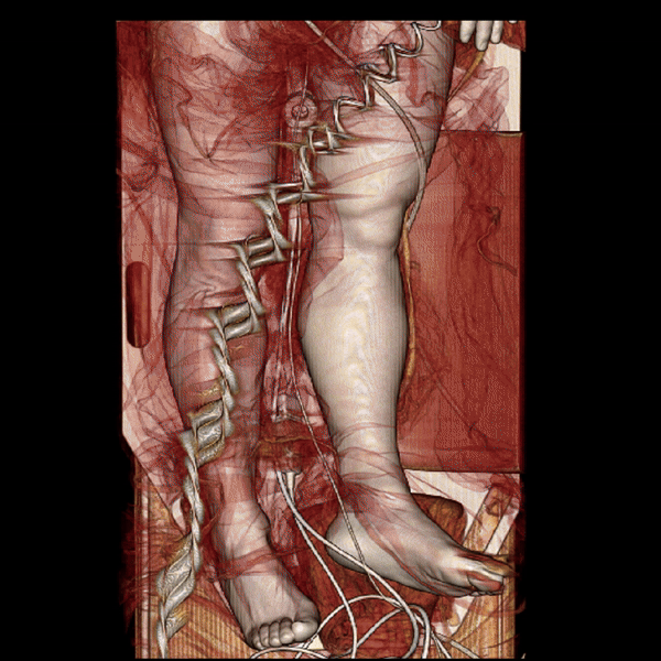
Figure B (Right): Rotational 3D reconstruction showing a brief view of the skin, muscle, and bone layers.

Below are the images the 3DQ Lab provided to the trauma team.

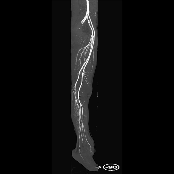
Figure C: The MIP clearly shows the lower extremity arteries on both sides, with no significant stenosis or obstructions, offering a clear view of blood flow from the pelvis to the legs.

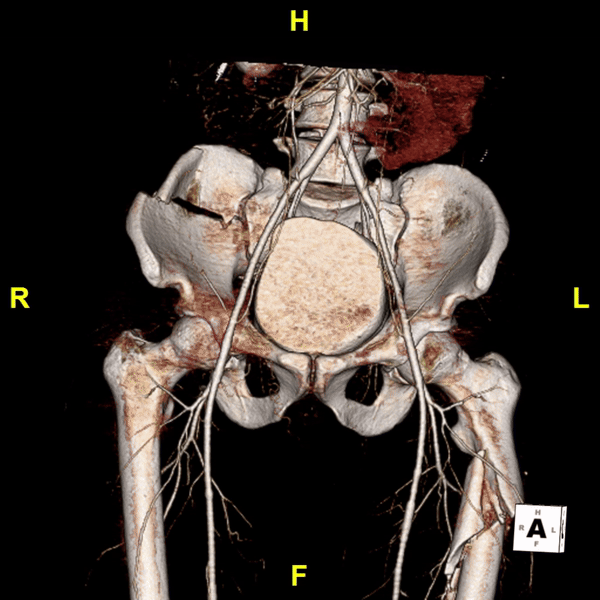
Figure D: A 3D reconstruction of the pelvis shows a comminuted fracture of the right iliac wing with extension into the sacroiliac joint, and nearby vascular structures.

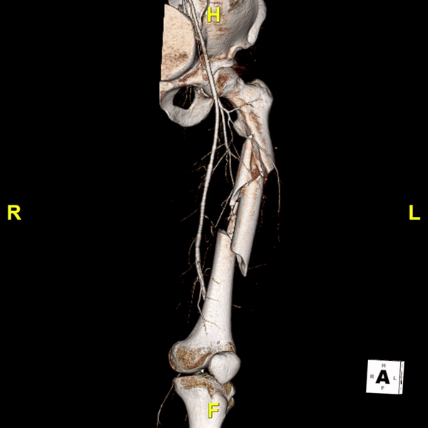
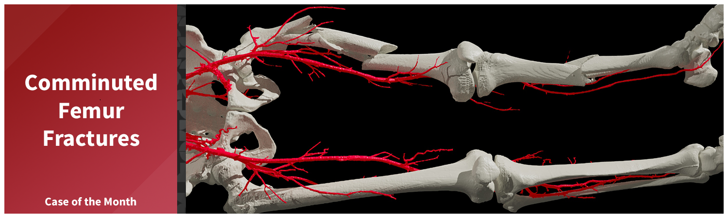
Figure E: A 3D reconstruction of the femur shows a severely comminuted fracture of the left femoral shaft with significant shortening, and nearby vascular structures.

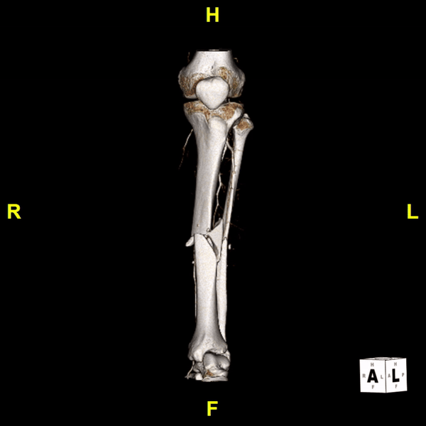
Figure F: A 3D reconstruction illustrating the severely comminuted fractures of the tibia and fibula, and nearby vascular structures.

Categories

  • Head & Neck
  • Chest
  • Abdomen & Pelvis
  • Upper Extremity
  • Lower Extremity
  • Case of the Month
  • Techniques

3DQ Lab – Grant Building

300 Pasteur Drive
Stanford, CA 94305

(650) 725-8432

Directions to Grant

3DQ Lab – Clark Building

318 Campus Drive
Stanford, CA 94305

(650) 725-6862

Directions to Clark

Learn More About the Lab

  • About Us
  • Meet the Team

Copyright © Stanford University

© Copyright - 3D and Quantitative Imaging Laboratory - Enfold Theme by Kriesi
  • LinkedIn
  • Instagram
Scaling and Sharing 3D Imaging SystemsCase of the Month – Watchman Device
Scroll to top