A patient with progressive neck pain and neurologic symptoms was referred to the 3DQ Lab for imaging due to concern for narrowing of the cervical spinal canal. Prior imaging demonstrated extensive ossification of the posterior longitudinal ligament from C2 through C6, resulting in spinal canal stenosis. Additional imaging was requested to better characterize the disease and support surgical planning.
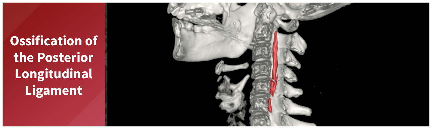
Ossification of the posterior longitudinal ligament, commonly referred to as OPLL, occurs when ligament tissue that is normally flexible gradually transforms into bone. When this occurs, it can reduce the space available for the spinal cord. Because the posterior longitudinal ligament lies directly in front of the cord, OPLL can have a significant impact on neurologic function.
Figure A (Right): Rotational 3D rendering of bone (white & transparent), and the ossified ligament (teal).
A CT scan of the cervical spine was obtained to further evaluate the presence and severity of OPLL. CT is well suited for this assessment because it clearly depicts bone and calcified structures, allowing the ossified ligament to be easily distinguished from surrounding soft tissues. The scan confirmed extensive OPLL from C2 through C6 with severe spinal canal stenosis.
Using the CT dataset, a radiologic technologist processed the images to create a series of advanced visualizations that highlight the distribution of ossification and its relationship to surrounding anatomy. These images provide different perspectives of the cervical spine and help translate cross-sectional findings into more intuitive views.
Figure B: 360-degree CPR view of the spinal canal. Learn more about CPRs here.
Figure C: 360-degree CPR view of the vertebral bodies.
Figure D: Grayscale rotational volume render of the spine.
Figure E: Grayscale rotational volume render of the spine.
Volume rendering and cutplane techniques were also used to create views that expose the interior of the spinal canal. These images show how the ossified posterior longitudinal ligament occupies space normally reserved for the spinal cord, offering a clearer sense of the degree of compression. All of these images were provided to the surgical team to add context and support presurgical planning by illustrating the spatial impact of the disease.
Figure F: Rotational volume render with a cutplane exposing the spinal canal. Learn more about cutplanes here.
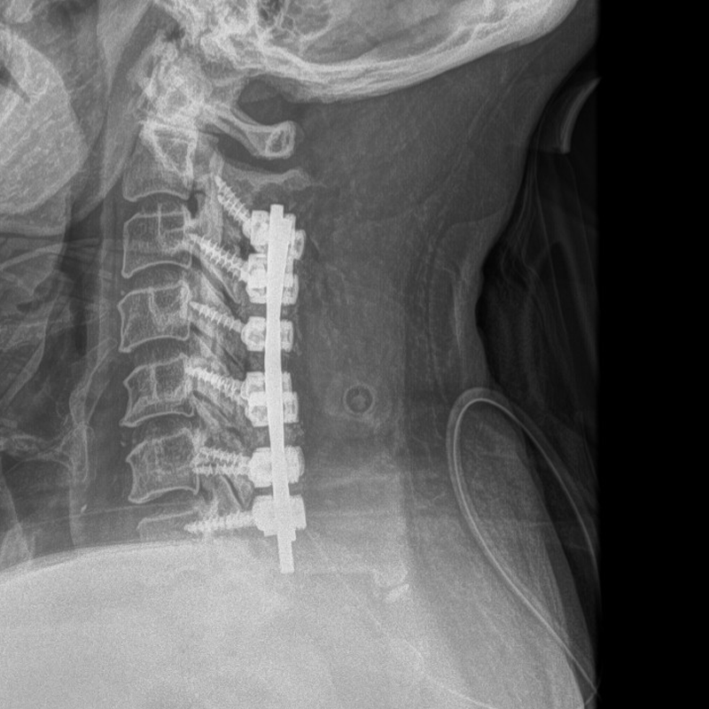
Given the severity of spinal canal stenosis and associated spinal cord compression, surgical intervention was pursued. Treatment for OPLL in cases such as this typically focuses on decompressing the spinal cord and stabilizing the cervical spine to prevent further neurologic decline. In this case, a postoperative X-ray demonstrates the implanted hardware used to provide spinal stability following decompression.
Figure G (Right): Post-op X-ray, showing implanted hardware.