3D and Quantitative Imaging Laboratory
  • Home
  • Education
    • Education
    • Educational Post Feed
  • Research
    • Research
    • Validation Cohort: Acute Uncomplicated Type-B Aortic Dissection
  • Patient Care
    • Patient Care
    • 3D Printing in Medicine
    • 3DQ Lab TRAC Team (Tumor Response)
  • Case Studies
    • Case Studies
    • Innovation Projects
  • Click to open the search input field Click to open the search input field Search
  • Menu Menu

February 2017 – Aortic Surveillance

Case of the Month, Chest, Techniques

Cross-Section:

VR Movie:
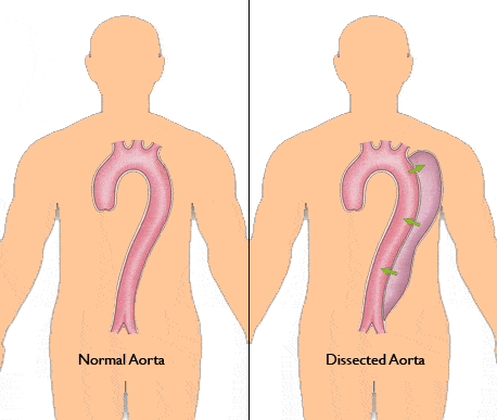
Figure A

Figure B

70 year-old male has an aortic dissection that begins in the Ascending Aorta and extends along the Aorta involving multiple abdominal and pelvic arteries along the course. The video with black/white image shows what is called a cross-section of the aorta from the heart, through the aorta, all the way to the pelvic vessels. A normal aorta will have a single-bordered, round/elliptical appearance (Figure A). This particular aorta has multiple borders (darkness in the round shape) showing a detached inner layer of the aorta (intima).

The 3DQ Lab at Stanford specializes at visualizing and quantifying patients with this condition. To do this, we standardized measurement locations and provide precise measurements at these landmarks for each aorta of interest; these landmarks are shown in Figure B. We currently track over 1,500 patients with Aorta issues such as dissection. Our standardized analysis approach provides a succinct graph plotting historic aorta measurements for each patient (Figure C).

For more information about dissections, please visit: http://www.iradonline.org/about.html

Shannon Walters, MS, RT(MR)
Executive Manager, 3D and Quantitative Imaging Laboratory

March 7, 2017/by cletrong
https://3dqlab.stanford.edu/wp-content/uploads/2017/03/nl_ds-aorta_L-1.gif 387 458 cletrong https://3dqlab.stanford.edu/wp-content/uploads/2023/08/3DQ-Website-Logo-Header3.png cletrong2017-03-07 14:57:092023-09-19 08:37:54February 2017 – Aortic Surveillance
Page 2 of 212

Categories

  • Head & Neck
  • Chest
  • Abdomen & Pelvis
  • Upper Extremity
  • Lower Extremity
  • Case of the Month
  • Techniques

3DQ Lab – Grant Building

300 Pasteur Drive
Stanford, CA 94305

(650) 725-8432

Directions to Grant

3DQ Lab – Clark Building

318 Campus Drive
Stanford, CA 94305

(650) 725-6862

Directions to Clark

Learn More About the Lab

  • About Us
  • Meet the Team
Search Search

Copyright © Stanford University

© Copyright - 3D and Quantitative Imaging Laboratory - Enfold Theme by Kriesi
  • Link to LinkedIn
  • Link to Instagram
Scroll to top Scroll to top Scroll to top