3D and Quantitative Imaging Laboratory
  • Home
  • Education
    • Education
    • Educational Post Feed
  • Research
    • Research
    • Validation Cohort: Acute Uncomplicated Type-B Aortic Dissection
  • Patient Care
    • Patient Care
    • 3D Printing in Medicine
    • 3DQ Lab TRAC Team (Tumor Response)
  • Case Studies
    • Case Studies
    • Innovation Projects
  • Click to open the search input field Click to open the search input field Search
  • Menu Menu

3D Printed Microsurgical Planning Models

Collaborators: Stanford Division of Plastic & Reconstructive Surgery

Complex microsurgical reconstruction depends on identifying small blood vessels and understanding how they travel through surrounding tissue. While CT angiography provides this information, interpreting vascular anatomy on 2D images alone can be challenging and may lead to changes in surgical plans once dissection begins.

This study evaluated whether patient-specific 3D printed models improved planning for breast reconstruction and other microsurgical procedures. Compared to CT angiography alone, use of 3D models reduced flap harvest time and eliminated changes between the preoperative plan and the flap ultimately performed.

Publication Link: Thieme

Figure A: Rotational 3D rendering of a patient-specific DIEP flap planning model. Custom design elements are incorporated to provide measurement references and help indicate the location of perforators.
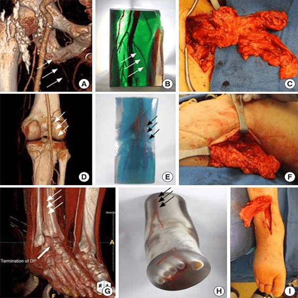
Figure B: Examples of planning models used for flap reconstruction. Each row shows a volume render, the corresponding 3D-printed model, and intraoperative photographs demonstrating how the models helped localize perforators and guide flap harvest.

The 3DQ Lab segmented the relevant anatomy from CT angiography, refined the models for printing, and produced multi-color PolyJet models that highlighted perforators and surrounding structures. These models were used during preoperative discussions and as intraoperative reference guides.

The study showed that physical models improved confidence in surgical planning while maintaining similar postoperative outcomes. Beyond breast reconstruction, the same approach was applied to burn reconstruction and lymphedema surgery, demonstrating how 3D printing can support complex microsurgical decision-making and education.

Categories

  • Head & Neck
  • Chest
  • Abdomen & Pelvis
  • Upper Extremity
  • Lower Extremity
  • Case of the Month
  • Techniques

3DQ Lab – Grant Building

300 Pasteur Drive
Stanford, CA 94305

(650) 725-8432

Directions to Grant

3DQ Lab – Clark Building

318 Campus Drive
Stanford, CA 94305

(650) 725-6862

Directions to Clark

Learn More About the Lab

  • About Us
  • Meet the Team
Search Search

Copyright © Stanford University

© Copyright - 3D and Quantitative Imaging Laboratory - Enfold Theme by Kriesi
  • Link to LinkedIn
  • Link to Instagram
Link to: Aortic Dissection Hemodynamic Modeling Link to: Aortic Dissection Hemodynamic Modeling Aortic Dissection Hemodynamic Modeling Link to: AI Modeling of Congenital Heart Disease Link to: AI Modeling of Congenital Heart Disease AI Modeling of Congenital Heart Disease
Scroll to top Scroll to top Scroll to top