3D and Quantitative Imaging Laboratory
  • Home
  • Education
    • Education
    • Educational Post Feed
  • Research
    • Research
    • Validation Cohort: Acute Uncomplicated Type-B Aortic Dissection
  • Patient Care
    • Patient Care
    • 3D Printing in Medicine
    • 3DQ Lab TRAC Team (Tumor Response)
  • Case Studies
    • Case Studies
    • Innovation Projects
  • Click to open the search input field Click to open the search input field Search
  • Menu Menu

AI Modeling of Congenital Heart Disease

Collaborators: Stanford Engineering Marsden Lab

Congenital heart disease includes a wide range of structural abnormalities, many of which are rare and highly variable from one patient to another. This diversity makes it difficult to assemble datasets large enough to train and validate artificial intelligence models, particularly for uncommon diagnoses such as Tetralogy of Fallot, transposition of the great arteries, and pulmonary atresia.

This study developed a deep learning approach to generate realistic 3D heart anatomies tailored to specific congenital heart disease diagnoses. By learning both the characteristic structural features of each condition and the shape variations seen across patients, the model was able to create synthetic anatomies that preserved clinically important abnormalities. These virtual hearts can be used to augment training datasets and create meshes for computational simulations.

Publication Link: Springer Nature
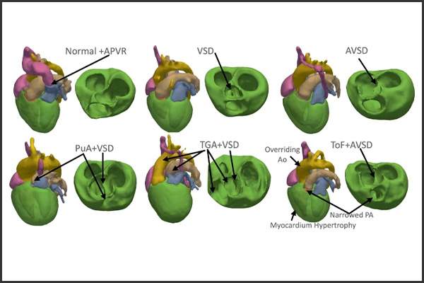
Figure A: AI-generated 3D heart models demonstrating how the network learned the characteristic anatomical features of different congenital heart disease diagnoses.
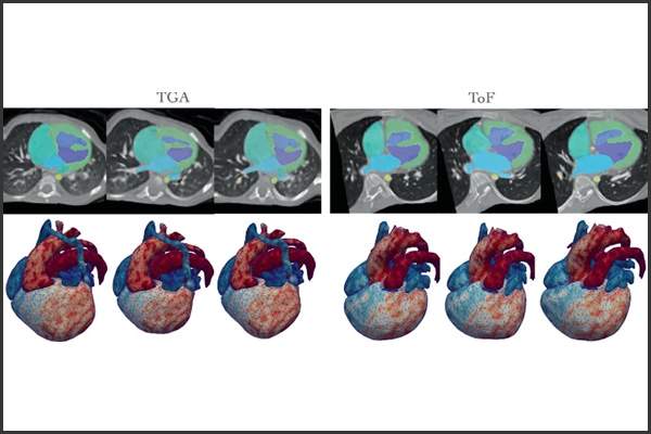
Figure B: AI-generated examples showing synthetic CT images with segmentation overlays and corresponding 3D heart models for different congenital heart disease anatomies.

The 3DQ Lab contributed expertise in cardiac segmentation and 3D modeling, helping prepare and refine the anatomical datasets used to train the model. This included reviewing segmentations for accuracy and ensuring that the resulting 3D representations preserved the structural features relevant to each congenital heart disease type.

The model successfully generated diagnosis-specific heart anatomies and reconstructed previously unseen cases more accurately than prior methods. This approach provides a scalable way to create synthetic datasets for rare congenital heart diseases, supporting future work in AI-based segmentation, simulation, and personalized treatment planning.

Categories

  • Head & Neck
  • Chest
  • Abdomen & Pelvis
  • Upper Extremity
  • Lower Extremity
  • Case of the Month
  • Techniques

3DQ Lab – Grant Building

300 Pasteur Drive
Stanford, CA 94305

(650) 725-8432

Directions to Grant

3DQ Lab – Clark Building

318 Campus Drive
Stanford, CA 94305

(650) 725-6862

Directions to Clark

Learn More About the Lab

  • About Us
  • Meet the Team
Search Search

Copyright © Stanford University

© Copyright - 3D and Quantitative Imaging Laboratory - Enfold Theme by Kriesi
  • Link to LinkedIn
  • Link to Instagram
Link to: Microsurgical Planning Models Link to: Microsurgical Planning Models Microsurgical Planning Models Link to: 3D Printing for Pediatric Transplant Patients Link to: 3D Printing for Pediatric Transplant Patients 3D Printing for Pediatric Transplant Patients
Scroll to top Scroll to top Scroll to top