 https://3dqlab.stanford.edu/wp-content/uploads/2025/05/diep300.png
300
300
Kyle Gifford
https://3dqlab.stanford.edu/wp-content/uploads/2023/08/3DQ-Website-Logo-Header3.png
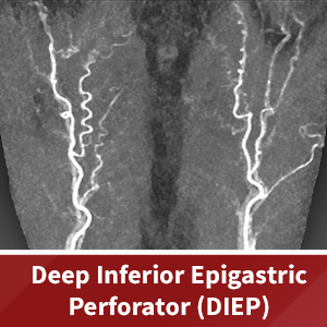
Kyle Gifford2025-05-27 10:36:282025-05-30 10:48:36DIEP (Deep Inferior Epigastric Perforator) Flap Reconstruction
https://3dqlab.stanford.edu/wp-content/uploads/2025/05/diep300.png
300
300
Kyle Gifford
https://3dqlab.stanford.edu/wp-content/uploads/2023/08/3DQ-Website-Logo-Header3.png
Kyle Gifford2025-05-27 10:36:282025-05-30 10:48:36DIEP (Deep Inferior Epigastric Perforator) Flap Reconstruction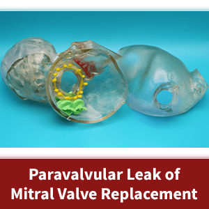 https://3dqlab.stanford.edu/wp-content/uploads/2025/04/pvl-300.png
300
300
Kyle Gifford
https://3dqlab.stanford.edu/wp-content/uploads/2023/08/3DQ-Website-Logo-Header3.png
Kyle Gifford2025-04-16 12:01:072025-05-05 15:44:48Case of the Month: Mitral Valve Replacement with Paravalvular Leak
https://3dqlab.stanford.edu/wp-content/uploads/2025/04/pvl-300.png
300
300
Kyle Gifford
https://3dqlab.stanford.edu/wp-content/uploads/2023/08/3DQ-Website-Logo-Header3.png
Kyle Gifford2025-04-16 12:01:072025-05-05 15:44:48Case of the Month: Mitral Valve Replacement with Paravalvular Leak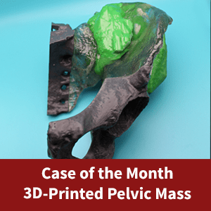 https://3dqlab.stanford.edu/wp-content/uploads/2025/01/1124-pelvic-mass-300.png
300
300
Kyle Gifford
https://3dqlab.stanford.edu/wp-content/uploads/2023/08/3DQ-Website-Logo-Header3.png
Kyle Gifford2025-01-06 09:29:022025-02-12 10:35:23Case of the Month – Pelvic Mass
https://3dqlab.stanford.edu/wp-content/uploads/2025/01/1124-pelvic-mass-300.png
300
300
Kyle Gifford
https://3dqlab.stanford.edu/wp-content/uploads/2023/08/3DQ-Website-Logo-Header3.png
Kyle Gifford2025-01-06 09:29:022025-02-12 10:35:23Case of the Month – Pelvic Mass https://3dqlab.stanford.edu/wp-content/uploads/2024/08/CoTM-Spine-Hardware-300.png
300
320
Kyle Gifford
https://3dqlab.stanford.edu/wp-content/uploads/2023/08/3DQ-Website-Logo-Header3.png
Kyle Gifford2024-08-27 16:32:472024-08-29 11:28:24Idiopathic Scoliosis
https://3dqlab.stanford.edu/wp-content/uploads/2024/08/CoTM-Spine-Hardware-300.png
300
320
Kyle Gifford
https://3dqlab.stanford.edu/wp-content/uploads/2023/08/3DQ-Website-Logo-Header3.png
Kyle Gifford2024-08-27 16:32:472024-08-29 11:28:24Idiopathic Scoliosis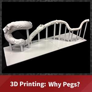 https://3dqlab.stanford.edu/wp-content/uploads/2024/06/pegs-300.jpg
300
300
Kyle Gifford
https://3dqlab.stanford.edu/wp-content/uploads/2023/08/3DQ-Website-Logo-Header3.png
Kyle Gifford2024-06-20 15:31:582024-06-20 15:32:003D Printing: Pegs
https://3dqlab.stanford.edu/wp-content/uploads/2024/06/pegs-300.jpg
300
300
Kyle Gifford
https://3dqlab.stanford.edu/wp-content/uploads/2023/08/3DQ-Website-Logo-Header3.png
Kyle Gifford2024-06-20 15:31:582024-06-20 15:32:003D Printing: Pegs https://3dqlab.stanford.edu/wp-content/uploads/2024/01/may-thurner-300.jpg
300
300
Kyle Gifford
https://3dqlab.stanford.edu/wp-content/uploads/2023/08/3DQ-Website-Logo-Header3.png
Kyle Gifford2024-01-26 13:19:592024-02-01 09:36:55May Thurner Syndrome
https://3dqlab.stanford.edu/wp-content/uploads/2024/01/may-thurner-300.jpg
300
300
Kyle Gifford
https://3dqlab.stanford.edu/wp-content/uploads/2023/08/3DQ-Website-Logo-Header3.png
Kyle Gifford2024-01-26 13:19:592024-02-01 09:36:55May Thurner Syndrome https://3dqlab.stanford.edu/wp-content/uploads/2023/02/Mitral-Valve-Repair-Title.png
300
300
Kyle Gifford
https://3dqlab.stanford.edu/wp-content/uploads/2023/08/3DQ-Website-Logo-Header3.png
Kyle Gifford2023-02-24 19:24:402023-03-27 06:59:193D Printing – Mitral Valve Repair
https://3dqlab.stanford.edu/wp-content/uploads/2023/02/Mitral-Valve-Repair-Title.png
300
300
Kyle Gifford
https://3dqlab.stanford.edu/wp-content/uploads/2023/08/3DQ-Website-Logo-Header3.png
Kyle Gifford2023-02-24 19:24:402023-03-27 06:59:193D Printing – Mitral Valve Repair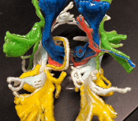 https://3dqlab.stanford.edu/wp-content/uploads/2022/12/Screen-Shot-2019-06-14-at-11.17.23-AM-1.png
556
458
shanwalt
https://3dqlab.stanford.edu/wp-content/uploads/2023/08/3DQ-Website-Logo-Header3.png
shanwalt2019-06-11 15:47:262023-09-19 08:22:13March 2019: 3D Printing Fiber Tracts in Brain
https://3dqlab.stanford.edu/wp-content/uploads/2022/12/Screen-Shot-2019-06-14-at-11.17.23-AM-1.png
556
458
shanwalt
https://3dqlab.stanford.edu/wp-content/uploads/2023/08/3DQ-Website-Logo-Header3.png
shanwalt2019-06-11 15:47:262023-09-19 08:22:13March 2019: 3D Printing Fiber Tracts in Brain https://3dqlab.stanford.edu/wp-content/uploads/2018/03/2-1024x770-1.png
602
800
cletrong
https://3dqlab.stanford.edu/wp-content/uploads/2023/08/3DQ-Website-Logo-Header3.png
cletrong2018-03-21 16:15:292023-09-19 08:29:483D Print of Cervical Spine Fracture
https://3dqlab.stanford.edu/wp-content/uploads/2018/03/2-1024x770-1.png
602
800
cletrong
https://3dqlab.stanford.edu/wp-content/uploads/2023/08/3DQ-Website-Logo-Header3.png
cletrong2018-03-21 16:15:292023-09-19 08:29:483D Print of Cervical Spine Fracture https://3dqlab.stanford.edu/wp-content/uploads/2023/08/3DQ-Website-Logo-Header3.png
0
0
Kyle Gifford
https://3dqlab.stanford.edu/wp-content/uploads/2023/08/3DQ-Website-Logo-Header3.png
Kyle Gifford2017-05-03 07:17:082023-09-19 08:36:26July 2017 – 3D Printed Brain & Electrodes
https://3dqlab.stanford.edu/wp-content/uploads/2023/08/3DQ-Website-Logo-Header3.png
0
0
Kyle Gifford
https://3dqlab.stanford.edu/wp-content/uploads/2023/08/3DQ-Website-Logo-Header3.png
Kyle Gifford2017-05-03 07:17:082023-09-19 08:36:26July 2017 – 3D Printed Brain & Electrodes https://3dqlab.stanford.edu/wp-content/uploads/2016/12/aortaarrow-1.png
600
800
cletrong
https://3dqlab.stanford.edu/wp-content/uploads/2023/08/3DQ-Website-Logo-Header3.png
cletrong2016-12-26 20:37:592023-09-19 08:38:46December 2016 – 3D Printed Aorta for TAVR
https://3dqlab.stanford.edu/wp-content/uploads/2016/12/aortaarrow-1.png
600
800
cletrong
https://3dqlab.stanford.edu/wp-content/uploads/2023/08/3DQ-Website-Logo-Header3.png
cletrong2016-12-26 20:37:592023-09-19 08:38:46December 2016 – 3D Printed Aorta for TAVR https://3dqlab.stanford.edu/wp-content/uploads/2016/04/Capture3-1-1.png
449
400
cletrong
https://3dqlab.stanford.edu/wp-content/uploads/2023/08/3DQ-Website-Logo-Header3.png
cletrong2016-04-01 21:09:172023-09-19 08:43:49March 2016
https://3dqlab.stanford.edu/wp-content/uploads/2015/02/1502COTM-Shannon-1-1.png
450
600
admin
https://3dqlab.stanford.edu/wp-content/uploads/2023/08/3DQ-Website-Logo-Header3.png
admin2015-02-28 22:22:112023-09-19 08:51:00February 2015
https://3dqlab.stanford.edu/wp-content/uploads/2016/04/Capture3-1-1.png
449
400
cletrong
https://3dqlab.stanford.edu/wp-content/uploads/2023/08/3DQ-Website-Logo-Header3.png
cletrong2016-04-01 21:09:172023-09-19 08:43:49March 2016
https://3dqlab.stanford.edu/wp-content/uploads/2015/02/1502COTM-Shannon-1-1.png
450
600
admin
https://3dqlab.stanford.edu/wp-content/uploads/2023/08/3DQ-Website-Logo-Header3.png
admin2015-02-28 22:22:112023-09-19 08:51:00February 2015