Augmented Reality for Kidney Donor Planning
Collaborators: Stanford Abdominal Transplantation
Preoperative understanding of kidney donor vascular anatomy is critical for selecting the appropriate organ and planning surgery. This is typically done using pre-calculated 2D volume-rendered images from CT or MR angiography, which limit interaction and may not fully convey complex spatial relationships.
In this work, a new rendering approach, NESTIS-VR, was developed to enable real-time, interactive 3D visualization on augmented reality headsets without requiring external computing hardware. Surgeons were able to adjust rendering parameters and explore anatomy from multiple viewpoints. Compared to standard 2D renderings, this approach significantly improved surgeon confidence in assessing renal arterial anatomy.
Publication Link: ScienceDirect
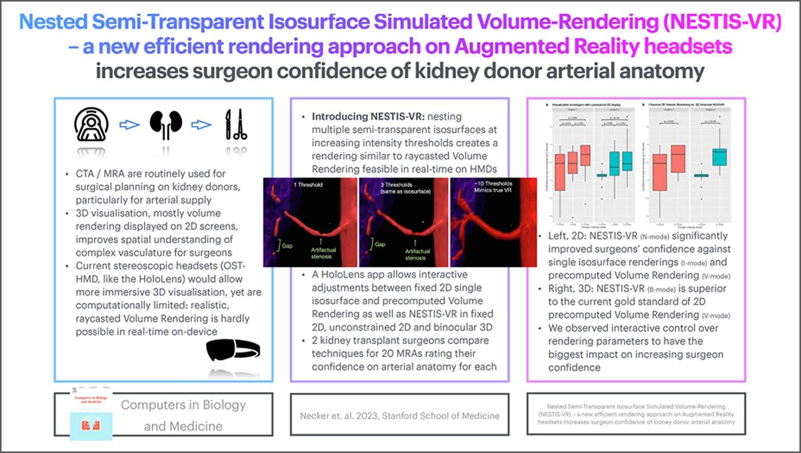
Figure A: Graphical abstract from the study illustrating the NESTIS-VR rendering approach and its impact on surgeon confidence in assessing kidney donor arterial anatomy.
The 3DQ Lab contributed by providing the patient-specific anatomical models used in this study. Segmentation of kidney vasculature and surrounding structures had already been performed as part of routine 3D imaging workflows, supporting tasks such as volume rendering and measurement. These segmentations were exported as aligned 3D mesh files and provided for use in the augmented reality system, forming the foundation for the interactive visualization.
By enabling direct interaction with patient-specific anatomy, this approach improves how surgeons interpret complex vascular structures before surgery. Increased confidence in anatomical assessment may support more informed donor selection and surgical planning, particularly in cases with complex vascular anatomy.
3DQ Lab – Grant Building
3DQ Lab – Clark Building
Learn More About the Lab
Copyright © Stanford University
