
3D Printed Microsurgical Planning Models
Complex microsurgical reconstruction depends on identifying small blood vessels and understanding how they travel through surrounding tissue. While CT angiography provides this information, interpreting vascular anatomy on 2D images alone can be challenging and may lead to changes in surgical plans once dissection begins.
This study evaluated whether patient-specific 3D printed models improved planning for breast reconstruction and other microsurgical procedures. Compared to CT angiography alone, use of 3D models reduced flap harvest time and eliminated changes between the preoperative plan and the flap ultimately performed.
Publication Link: Thieme
Figure A: Rotational 3D rendering of a patient-specific DIEP flap planning model. Custom design elements are incorporated to provide measurement references and help indicate the location of perforators.
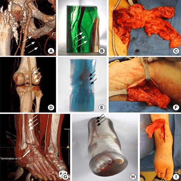
Figure B: Examples of planning models used for flap reconstruction. Each row shows a volume render, the corresponding 3D-printed model, and intraoperative photographs demonstrating how the models helped localize perforators and guide flap harvest.