3D and Quantitative Imaging Laboratory
  • Home
  • Education
    • Education
    • Educational Post Feed
  • Research
    • Research
    • Validation Cohort: Acute Uncomplicated Type-B Aortic Dissection
  • Patient Care
    • Patient Care
    • 3D Printing in Medicine
    • 3DQ Lab TRAC Team (Tumor Response)
  • Case Studies
    • Case Studies
    • Innovation Projects
  • Click to open the search input field Click to open the search input field Search
  • Menu Menu

Prostate Needle Biopsy Grid

Collaborators: Stanford Urology

During transperineal prostate biopsy and cryoablation, needles are inserted through the perineum into specific locations within the prostate. Their position determines where tissue samples are obtained or what portions of tissue are frozen, making accurate entry points and angles essential for both diagnosis and treatment.

The Department of Urology developed the initial concept for a reusable guidance grid to help standardize needle placement. The device contains a pattern of openings that supports the needle as it is advanced. Interchangeable attachments allow the system to work with different procedural tools and accommodate variations in patient size and anatomy.

Figure A: 3D rendering of the current prostate needle guidance grid design, showing the indexed hole pattern and modular geometry used to support precise needle placement.
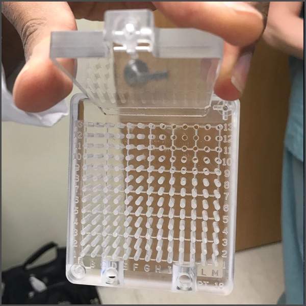
Figure B: Earlier prototype of the 3D printed prostate needle guidance grid with an attached procedural accessory.

Building on this concept, the 3DQ Lab took over the design and prototyping process to develop a clinically usable device. Drawing on expertise in 3D printing design, the lab refined the geometry, improved fit and durability, and developed interchangeable attachments. The 3DQ Lab 3D prints the grid and components and continues to provide  and update replacement parts as the design evolves.

This project demonstrates how clinician-engineer collaboration can transform a rough concept into a durable procedural tool. The guidance grid has supported more than six years of ongoing clinical use, helping improve needle stability and procedural consistency during transperineal prostate interventions.

Categories

  • Head & Neck
  • Chest
  • Abdomen & Pelvis
  • Upper Extremity
  • Lower Extremity
  • Case of the Month
  • Techniques

3DQ Lab – Grant Building

300 Pasteur Drive
Stanford, CA 94305

(650) 725-8432

Directions to Grant

3DQ Lab – Clark Building

318 Campus Drive
Stanford, CA 94305

(650) 725-6862

Directions to Clark

Learn More About the Lab

  • About Us
  • Meet the Team
Search Search

Copyright © Stanford University

© Copyright - 3D and Quantitative Imaging Laboratory - Enfold Theme by Kriesi
  • Link to LinkedIn
  • Link to Instagram
Link to: Silicone Heart Models for Septation Planning Link to: Silicone Heart Models for Septation Planning Silicone Heart Models for Septation Planning Link to: Custom Foot Orthotic Design Link to: Custom Foot Orthotic Design Custom Foot Orthotic Design
Scroll to top Scroll to top Scroll to top