3D and Quantitative Imaging Laboratory
  • Home
  • Education
    • Education
    • Educational Post Feed
  • Research
    • Research
    • Validation Cohort: Acute Uncomplicated Type-B Aortic Dissection
  • Patient Care
    • Patient Care
    • 3D Printing in Medicine
    • 3DQ Lab TRAC Team (Tumor Response)
  • Case Studies
    • Case Studies
    • Innovation Projects
  • Click to open the search input field Click to open the search input field Search
  • Menu Menu

Silicone Heart Models for Septation Planning

Collaborators: Stanford Cardiovascular Engineering Research Lab

In some congenital heart defects, the heart’s chambers and blood vessels are arranged in ways that make it difficult to predict how blood flow will change after surgery. For procedures like septation repair, clinicians may want to evaluate how a proposed reconstruction will affect circulation before entering the operating room.

This project combined computational modeling and ex vivo testing to study surgical repair on a patient-specific basis. Using CT, MRI, and catheterization data, cardiology teams developed computer simulations and mock circulatory loop experiments to compare blood flow before and after repair.

Figure A: Exploded view of the four-piece 3D printed mold designed to cast a patient-specific silicone heart model. Integrated pouring and vent holes allow silicone to fill the mold while enabling the cast to be removed without damaging the final model.
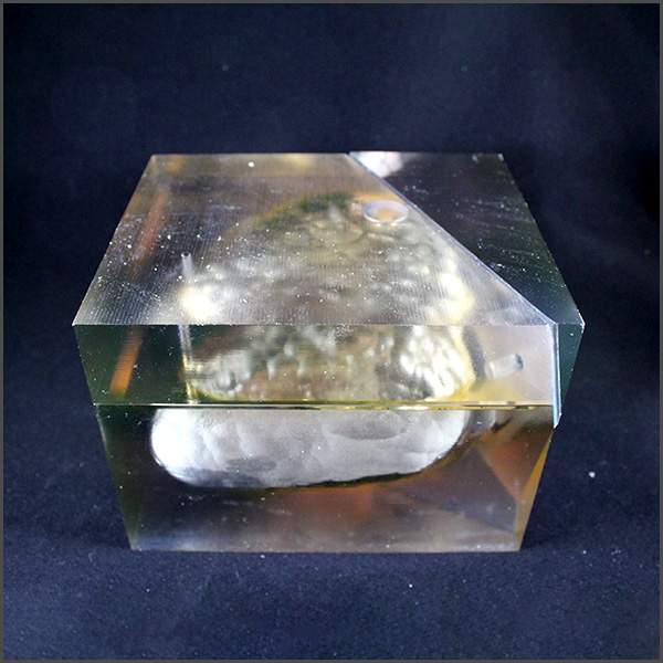
Figure B: Fully assembled 3D printed mold used to cast the silicone heart model.

The 3DQ Lab created patient-specific cardiac segmentations and designed and 3D printed four molds used to cast silicone heart models. These flexible models were placed in a mock circulatory loop, allowing investigators to measure how different repair strategies affected blood flow under controlled conditions.

By combining virtual simulations with physical testing, this approach provided a way to evaluate surgical strategies before intervention. The project demonstrates how patient-specific modeling and 3D printing can support treatment planning and advance research in complex congenital heart disease.

Categories

  • Head & Neck
  • Chest
  • Abdomen & Pelvis
  • Upper Extremity
  • Lower Extremity
  • Case of the Month
  • Techniques

3DQ Lab – Grant Building

300 Pasteur Drive
Stanford, CA 94305

(650) 725-8432

Directions to Grant

3DQ Lab – Clark Building

318 Campus Drive
Stanford, CA 94305

(650) 725-6862

Directions to Clark

Learn More About the Lab

  • About Us
  • Meet the Team
Search Search

Copyright © Stanford University

© Copyright - 3D and Quantitative Imaging Laboratory - Enfold Theme by Kriesi
  • Link to LinkedIn
  • Link to Instagram
Link to: 4D Flow MRI for CTEPH Link to: 4D Flow MRI for CTEPH 4D Flow MRI for CTEPH Link to: Prostate Needle Biopsy Grid Link to: Prostate Needle Biopsy Grid Prostate Needle Biopsy Grid
Scroll to top Scroll to top Scroll to top