3D and Quantitative Imaging Laboratory
  • Home
  • Education
    • Education
    • Educational Post Feed
  • Research
    • Research
    • Validation Cohort: Acute Uncomplicated Type-B Aortic Dissection
  • Patient Care
    • Patient Care
    • 3D Printing in Medicine
    • 3DQ Lab TRAC Team (Tumor Response)
  • Case Studies
    • Case Studies
    • Innovation Projects
  • Click to open the search input field Click to open the search input field Search
  • Menu Menu

Omental Flap Reconstruction

Collaborators: Stanford Division of Plastic & Reconstructive Surgery

The omentum is a fatty apron-like structure within the abdomen that can be transferred as a vascularized flap for breast reconstruction. Stanford surgeons developed a novel technique that combines laparoscopic omental harvest with fat grafting to create a customized breast mound for patients who may not be candidates for more traditional reconstruction options.

In 2020, the Division of Plastic and Reconstructive Surgery approached the 3DQ Lab to explore whether CT imaging could be used to quantify breast volume before surgery. By measuring the breast with and without the overlying skin, the team could better estimate the amount of soft tissue required for reconstruction and compare that target volume with the tissue available from the omental flap.
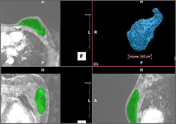
Figure A: Segmentation of the breast volume used for analysis, shown in cross-sectional CT images and segmentation (green) and as a volume render with calculated volume.
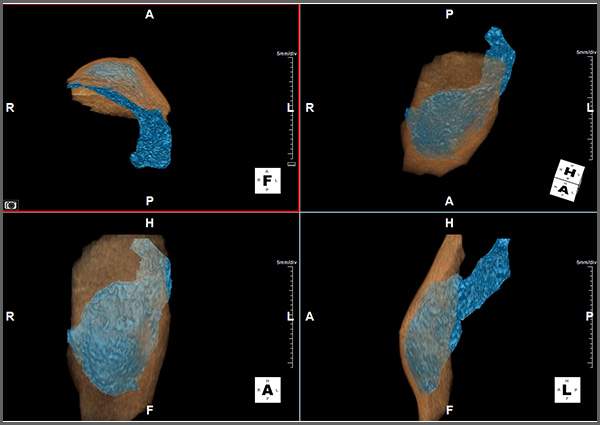
Figure B: Comparison of the segmented breast volume (blue) within the surrounding breast envelope (tan), used to estimate tissue requirements for omental flap reconstruction.

Working together, the surgical team and the 3DQ Lab developed a segmentation workflow to create 3D models and calculate these breast volumes from CT imaging. The resulting measurements provided objective volumetric data that supported publication and further evaluation of the reconstructive technique.

Stanford’s experience with omental flap reconstruction has since attracted visiting surgeons from across the United States and abroad who traveled to Stanford to observe and learn the procedure. This project illustrates how collaboration between Plastic and Reconstructive Surgery and the 3DQ Lab helped generate quantitative data that supported both academic publication and dissemination of a novel surgical approach.

Publication: PubMed

Categories

  • Head & Neck
  • Chest
  • Abdomen & Pelvis
  • Upper Extremity
  • Lower Extremity
  • Case of the Month
  • Techniques

3DQ Lab – Grant Building

300 Pasteur Drive
Stanford, CA 94305

(650) 725-8432

Directions to Grant

3DQ Lab – Clark Building

318 Campus Drive
Stanford, CA 94305

(650) 725-6862

Directions to Clark

Learn More About the Lab

  • About Us
  • Meet the Team
Search Search

Copyright © Stanford University

© Copyright - 3D and Quantitative Imaging Laboratory - Enfold Theme by Kriesi
  • Link to LinkedIn
  • Link to Instagram
Link to: Siemens Auto Stitch & Batch Link to: Siemens Auto Stitch & Batch Siemens Auto Stitch & Batch Link to: Speaking Engagements Link to: Speaking Engagements Speaking Engagements
Scroll to top Scroll to top Scroll to top