3D and Quantitative Imaging Laboratory
  • Home
  • Education
    • Education
    • Educational Post Feed
  • Research
    • Research
    • Validation Cohort: Acute Uncomplicated Type-B Aortic Dissection
  • Patient Care
    • Patient Care
    • 3D Printing in Medicine
    • 3DQ Lab TRAC Team (Tumor Response)
  • Case Studies
    • Case Studies
    • Innovation Projects
  • Click to open the search input field Click to open the search input field Search
  • Menu Menu

Essential Tremor Tract Mapping

Collaborators: Stanford Neuroimaging & Neurointervention (Neuroradiology)

Essential tremor is a neurologic disorder that causes rhythmic shaking with movement, most often in the hands, and it can interfere with eating, writing, and other daily tasks. One treatment option is MRI-guided focused ultrasound thalamotomy, which uses focused ultrasound energy to heat and destroy a small target in the brain without an incision. Targeting is typically planned in the ventral intermediate nucleus (VIM) of the thalamus on the side opposite the patient’s more affected hand.

Procedure planning can benefit from patient-specific “nerve pathway” mapping using diffusion MRI. A key pathway for tremor targeting is the dentato-rubro-thalamic tract (DRTT), which connects the cerebellum to the thalamus and helps regulate motor control. Stanford clinicians identified a workflow gap: there was no reliable internal method to generate and review these diffusion-based pathway maps in a standardized way for focused ultrasound planning.

Neuroradiology asked the Stanford 3DQ Lab to build a repeatable workflow that could produce these pathway maps and support the focused ultrasound planning and guidance process. A 3DQ Lab technologist attended an external conference to learn tract targeting approaches used by other institutions and field leaders, then worked with Stanford neuroradiologists to adapt the method to local imaging and review needs.
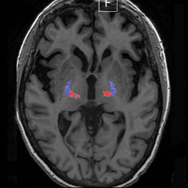
Figure A. Axial MRI with diffusion tractography overlay used for focused ultrasound planning, showing the dentato-rubro-thalamic tract (DRTT, red) and pyramidal (corticospinal) tract (blue) near the thalamus.
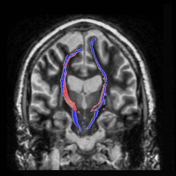
Figure B. Coronal MRI with diffusion tractography overlay showing the course of the DRTT (red) relative to the pyramidal (corticospinal) tract (blue) for pathway review during focused ultrasound planning.

The 3DQ Lab processed diffusion MRI data to generate tractography maps, then filtered large numbers of candidate fiber tracks down to the DRTT and displayed it alongside the pyramidal (corticospinal) tract, a nearby motor pathway used as a safety reference. Early cases required iteration to confirm anatomy, set consistent start and end regions, and correct registration errors that shifted pathway position relative to the MRI.

Diffusion scan settings also limited how clearly the pathway could be seen. The 3DQ Lab recommended updated scan parameters to improve pathway visibility and the stability of the tractography output. The resulting workflow produced standardized pathway overlays and review images that supported focused ultrasound targeting review and safety checks prior to ablation.

Categories

  • Head & Neck
  • Chest
  • Abdomen & Pelvis
  • Upper Extremity
  • Lower Extremity
  • Case of the Month
  • Techniques

3DQ Lab – Grant Building

300 Pasteur Drive
Stanford, CA 94305

(650) 725-8432

Directions to Grant

3DQ Lab – Clark Building

318 Campus Drive
Stanford, CA 94305

(650) 725-6862

Directions to Clark

Learn More About the Lab

  • About Us
  • Meet the Team
Search Search

Copyright © Stanford University

© Copyright - 3D and Quantitative Imaging Laboratory - Enfold Theme by Kriesi
  • Link to LinkedIn
  • Link to Instagram
Link to: TRAC Program Link to: TRAC Program TRAC Program Link to: Aortic Surveillance of Loeys Dietz Syndrome Link to: Aortic Surveillance of Loeys Dietz Syndrome Aortic Surveillance of Loeys Dietz Syndrome
Scroll to top Scroll to top Scroll to top