3D and Quantitative Imaging Laboratory
  • Home
  • Education
    • Education
    • Educational Post Feed
  • Research
    • Research
    • Validation Cohort: Acute Uncomplicated Type-B Aortic Dissection
  • Patient Care
    • Patient Care
    • 3D Printing in Medicine
    • 3DQ Lab TRAC Team (Tumor Response)
  • Case Studies
    • Case Studies
    • Innovation Projects
  • Click to open the search input field Click to open the search input field Search
  • Menu Menu

Aortic Surveillance Workflow

Collaborators: Stanford Cardiovascular Surgery

Patients with aortic aneurysms and dissections often undergo CT or MRI over many years to monitor changes in aortic size. Because surgical decisions may depend on small changes in diameter, measurements must be performed consistently across time, including when prior studies originate from outside institutions using different imaging protocols.

Before the 3DQ Lab became involved, Cardiovascular Surgery manually tracked measurements in spreadsheets and assembled graphs in PowerPoint. Communication relied heavily on email, and hundreds of outside studies had accumulated on disk without being fully reviewed or measured. This approach was labor intensive and made it difficult to provide timely information for clinic.

Figure A: Workflow diagram illustrating the decision-making process used to receive, process, and report Cardiovascular Surgery imaging requests.

Figure B: Automatically generated longitudinal graph showing aortic diameters at standardized locations across multiple imaging studies.
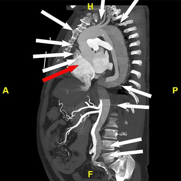
Figure C: Standardized measurement locations used throughout the aorta to ensure consistent comparison across current and prior examinations.

Since 2013, the 3DQ Lab has partnered with Stanford Cardiothoracic  Surgery to build a dedicated service infrastructure for measuring and reporting this concept of aortic surveillance. Standardized  measurements were defined at standardized  locations in the aorta. Any follow-up studies are measured at the same locations as prior imaging. A custom web portal was developed to request studies, prioritize urgent cases, track case status, and deliver results through a single shared interface.

Measurement values are stored in a structured database rather than spreadsheets. The system generates longitudinal reports on demand that include aortic diameters over time. Supporting the graphs are the underlying measurement values and annotated images archived in PACS. These reports are viewed online or downloaded as PDF summaries, providing the Cardiothoracic Surgery team on-demand access to the full measurement history.

The workflow also supports rapid turnaround for prioritized cases, allowing surgeons to review updated measurements immediately before a  patient visit – which occasionally occurs just hours after the imaging exam to be evaluated. Every measurement undergoes quality assurance by an experienced technologist to verify image quality, data integrity, and adherence to standardized measurement protocols.

Categories

  • Head & Neck
  • Chest
  • Abdomen & Pelvis
  • Upper Extremity
  • Lower Extremity
  • Case of the Month
  • Techniques

3DQ Lab – Grant Building

300 Pasteur Drive
Stanford, CA 94305

(650) 725-8432

Directions to Grant

3DQ Lab – Clark Building

318 Campus Drive
Stanford, CA 94305

(650) 725-6862

Directions to Clark

Learn More About the Lab

  • About Us
  • Meet the Team
Search Search

Copyright © Stanford University

© Copyright - 3D and Quantitative Imaging Laboratory - Enfold Theme by Kriesi
  • Link to LinkedIn
  • Link to Instagram
Link to: Automated Extraction of Measurements Link to: Automated Extraction of Measurements Automated Extraction of Measurements Link to: Non-Clinical Time Link to: Non-Clinical Time Non-Clinical Time
Scroll to top Scroll to top Scroll to top