3D and Quantitative Imaging Laboratory
  • Home
  • Education
    • Education
    • Educational Post Feed
  • Research
    • Research
    • Validation Cohort: Acute Uncomplicated Type-B Aortic Dissection
  • Patient Care
    • Patient Care
    • 3D Printing in Medicine
    • 3DQ Lab TRAC Team (Tumor Response)
  • Case Studies
    • Case Studies
    • Innovation Projects
  • Click to open the search input field Click to open the search input field Search
  • Menu Menu

Augmented Reality for Kidney Donor Planning

Collaborators: Stanford Abdominal Transplantation

Preoperative understanding of kidney donor vascular anatomy is critical for organ selection and surgical planning. This assessment is typically based on 2D volume-rendered images from CT or MR angiography, which limit interaction and may not fully convey complex spatial relationships.

A new rendering approach, NESTIS-VR, was developed to enable real-time, interactive 3D visualization on standalone augmented reality (AR) headsets. Surgeons were able to adjust rendering parameters and explore anatomy from multiple viewpoints. Compared to standard 2D renderings, this approach significantly improved surgeon confidence in assessing renal arterial anatomy.

Publication Link: ScienceDirect
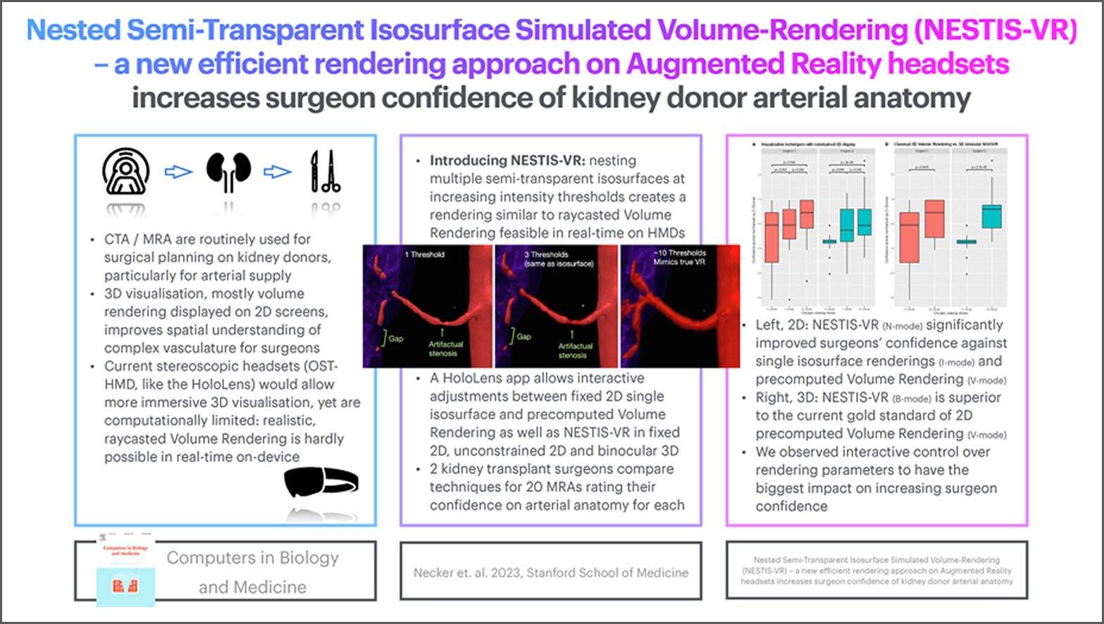
Figure A: Graphical abstract illustrating the NESTIS-VR approach and its impact on surgeon confidence in assessing kidney donor arterial anatomy.

The 3DQ Lab contributed the patient-specific anatomical models used in this study. Segmentations of kidney vasculature and surrounding structures were generated through routine 3D imaging workflows that support volume rendering and measurement. These segmentations were exported as aligned 3D mesh files and provided for use in the AR system, forming the foundation for the interactive visualization.

By enabling direct interaction with patient-specific anatomy, this approach improves how surgeons interpret complex vascular structures before surgery. Increased confidence in anatomical assessment may support better-informed donor selection and surgical planning, particularly in cases with complex vascular anatomy.

Categories

  • Head & Neck
  • Chest
  • Abdomen & Pelvis
  • Upper Extremity
  • Lower Extremity
  • Case of the Month
  • Techniques

3DQ Lab – Grant Building

300 Pasteur Drive
Stanford, CA 94305

(650) 725-8432

Directions to Grant

3DQ Lab – Clark Building

318 Campus Drive
Stanford, CA 94305

(650) 725-6862

Directions to Clark

Learn More About the Lab

  • About Us
  • Meet the Team
Search Search

Copyright © Stanford University

© Copyright - 3D and Quantitative Imaging Laboratory - Enfold Theme by Kriesi
  • Link to LinkedIn
  • Link to Instagram
Link to: 3D Liver Modeling for Pediatric Transplant Link to: 3D Liver Modeling for Pediatric Transplant 3D Liver Modeling for Pediatric Transplant Link to: Early Detection of Transplant-Related Lung Injury with CT Link to: Early Detection of Transplant-Related Lung Injury with CT Early Detection of Transplant-Related Lung Injury with CT
Scroll to top Scroll to top Scroll to top