3D and Quantitative Imaging Laboratory
  • Home
  • Education
    • Education
    • Educational Post Feed
  • Research
    • Research
    • Validation Cohort: Acute Uncomplicated Type-B Aortic Dissection
  • Patient Care
    • Patient Care
    • 3D Printing in Medicine
    • 3DQ Lab TRAC Team (Tumor Response)
  • Case Studies
    • Case Studies
    • Innovation Projects
  • Click to open the search input field Click to open the search input field Search
  • Menu Menu

Cumulus Breast Density

Collaborators: Stanford Epidemiology

Assessing breast cancer risk using mammography often includes evaluating breast density, but this has traditionally been studied using older film-based images. Today, most imaging is digital, and the images used in clinical care are processed for display. These processing steps can change how the image looks, raising questions about whether density measurements from these images are still reliable.

In this work, processed digital mammograms were used to measure breast density and evaluate its relationship with breast cancer risk. Density was assessed using Cumulus, a widely used tool for quantifying dense tissue on mammograms. The results showed that higher density remained strongly associated with increased risk, consistent with prior findings from film-based imaging. This supports the idea that routinely acquired clinical images can still provide meaningful and reproducible measurements.

Publication Link: PubMed
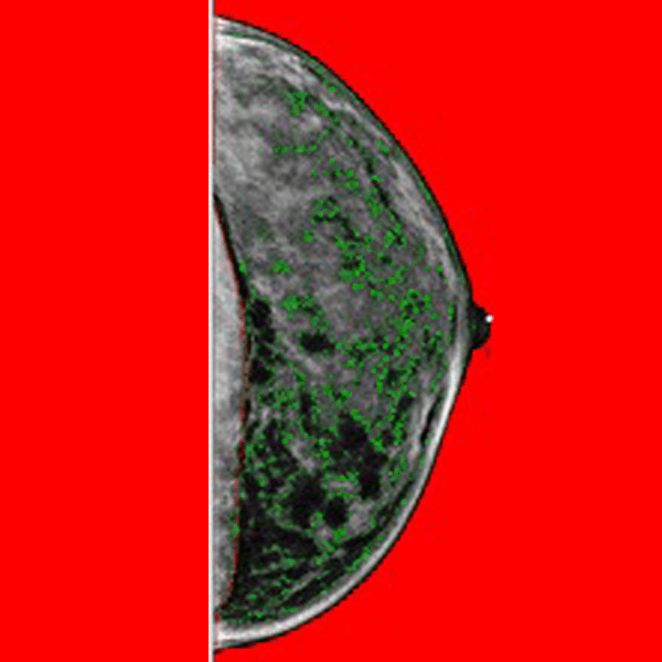
Figure A: Representative full-field digital mammography image prior to processing. This original image reflects the native acquisition resolution (70 micron pixel size) before any downsampling or filtering is applied.
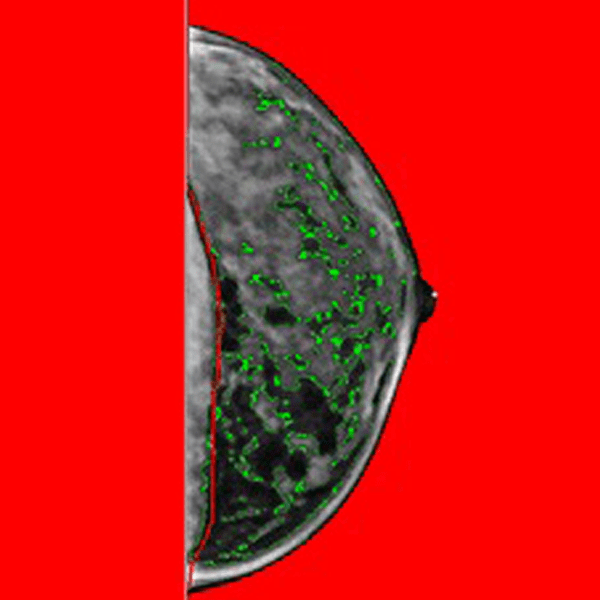
Figure B: Processed full-field digital mammography image after downsampling (to 200 micron pixel size) and median filtering (radius of three pixels), with dense tissue segmentation generated using Cumulus (outlined in green). These preprocessing steps support more consistent density assessment.

The 3DQ Lab contributed by supporting the scale needed for this type of analysis during the early adoption of digital mammography, with work spanning 2012 to 2014. A dataset of over 30,000 mammograms was processed using a standardized thresholding approach to extract density-related information. In parallel, database connectivity and a structured data workflow were developed to support consistent data handling across large volumes of imaging.

This matters for clinical workflows because it confirms that the images already used in routine care can support meaningful risk assessment. There is no need for additional image types or special data access, making it easier to use existing imaging data for both patient care and large-scale research.

Categories

  • Head & Neck
  • Chest
  • Abdomen & Pelvis
  • Upper Extremity
  • Lower Extremity
  • Case of the Month
  • Techniques

3DQ Lab – Grant Building

300 Pasteur Drive
Stanford, CA 94305

(650) 725-8432

Directions to Grant

3DQ Lab – Clark Building

318 Campus Drive
Stanford, CA 94305

(650) 725-6862

Directions to Clark

Learn More About the Lab

  • About Us
  • Meet the Team
Search Search

Copyright © Stanford University

© Copyright - 3D and Quantitative Imaging Laboratory - Enfold Theme by Kriesi
  • Link to LinkedIn
  • Link to Instagram
Link to: MRI-Aligned Prostate Cutting Guides Link to: MRI-Aligned Prostate Cutting Guides MRI-Aligned Prostate Cutting Guides Link to: Cerebral Aneurysm Growth and Hemodynamics Link to: Cerebral Aneurysm Growth and Hemodynamics Cerebral Aneurysm Growth and Hemodynamics
Scroll to top Scroll to top Scroll to top