
Cerebral Aneurysm Growth and Hemodynamics
Understanding why some cerebral aneurysms grow while others remain stable is a key challenge in neurovascular care. Blood flow dynamics are thought to play a role, but the specific hemodynamic patterns that drive growth are not fully defined. Measurements like wall shear stress and flow oscillation can vary widely depending on both anatomy and modeling approach, making it difficult to identify consistent predictors.
In this study, matched pairs of aneurysms with similar size and location were analyzed using computational fluid dynamics and finite element simulation to compare blood flow environments between growing and stable cases. These simulations estimated blood flow patterns from existing vascular CT datasets, allowing researchers to derive functional flow information from anatomical imaging without requiring additional scans or invasive procedures.
Several flow-related metrics were evaluated, with particular attention to regions exposed to lower wall shear stress. The results showed that growing aneurysms tended to have a larger portion of their surface exposed to low shear conditions, supporting prior observations that altered blood flow environments may contribute to aneurysm progression.
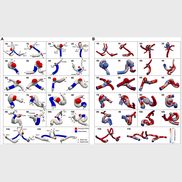
Figure A: Eleven matched pairs of cerebral aneurysms, with growing (G) and stable (S) cases. The aneurysm dome is shown in red and the parent artery in blue.
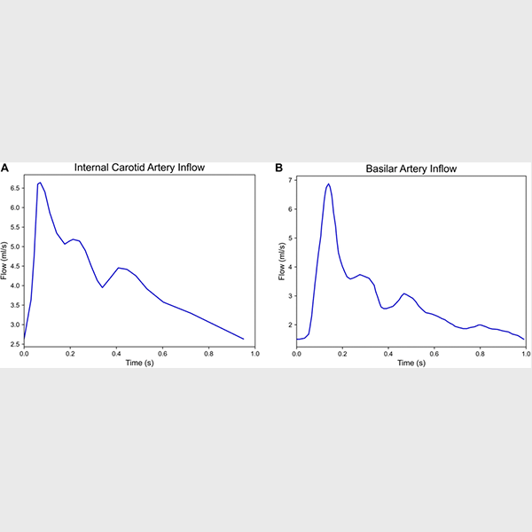
Figure B: Flow waveforms applied at model inlets, including the internal carotid artery (ICA) and basilar artery (BA).
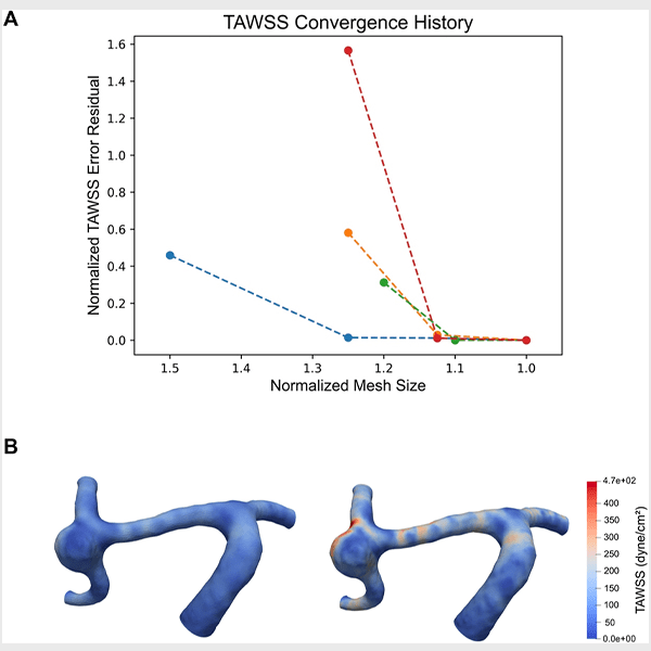
Figure C: Time-averaged wall shear stress (TAWSS) showing how results change with increasing simulation detail.