
Fluorescence Tumor Imaging
Accurately identifying tumor margins during brain tumor surgery is challenging because current imaging methods do not always clearly distinguish tumor from normal tissue. In high-grade gliomas, this is especially important, as even small amounts of residual tumor can impact patient outcomes. While standard imaging and navigation tools provide guidance, they often lack the specificity needed to visualize infiltrative tumor beyond the obvious boundaries. In this work, a targeted fluorescence imaging approach using panitumumab-IRDye800 was evaluated to improve intraoperative visualization by highlighting tumor tissue in real time based on molecular expression.
This approach uses an antibody that targets the epidermal growth factor receptor, which is commonly overexpressed in high-grade gliomas. Patients received the agent prior to surgery, allowing it to accumulate in tumor tissue. During surgery, near-infrared imaging was used to visualize fluorescence, helping differentiate tumor from the surrounding brain. The study demonstrated that this method can improve contrast compared to standard imaging, including detection of tumor beyond the contrast-enhancing regions seen on MRI. Tumor tissue showed strong fluorescence, while normal brain tissue showed minimal signal, supporting its ability to better define tumor boundaries.
Publication Link: Theranostics
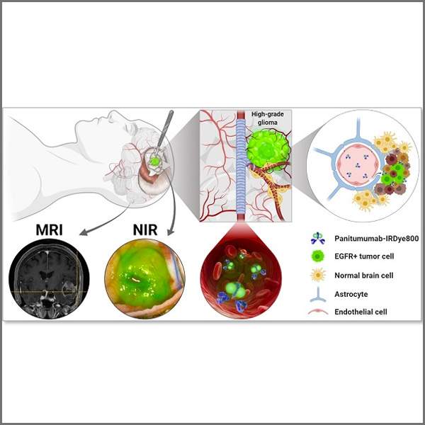
Figure A: Overview of fluorescence-guided glioma surgery, combining MRI and near-infrared imaging to help visualize tumor tissue during resection.
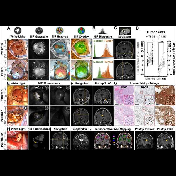
Figure B: Imaging overlays combining fluorescence imaging with MRI and intraoperative functional MRI mapping, supported by imaging workflows from the 3DQ Lab.