3D and Quantitative Imaging Laboratory
  • Home
  • Education
    • Education
    • Educational Post Feed
  • Research
    • Research
    • Validation Cohort: Acute Uncomplicated Type-B Aortic Dissection
  • Patient Care
    • Patient Care
    • 3D Printing in Medicine
    • 3DQ Lab TRAC Team (Tumor Response)
  • Case Studies
    • Case Studies
    • Innovation Projects
  • Click to open the search input field Click to open the search input field Search
  • Menu Menu

Predicting Bypass Graft Failure

Collaborators: Marsden Lab

Coronary artery bypass grafting (CABG) is widely used to treat advanced coronary artery disease, but long-term success depends on the durability of the grafts. Saphenous vein grafts are commonly used, yet many develop narrowing or occlusion over time. These changes are often silent and difficult to predict using standard clinical tools.

In this work, patient-specific models derived from CT angiography were used to investigate how blood flow contributes to graft failure. Computational fluid dynamics (CFD) simulations were performed to evaluate hemodynamic forces within each graft, with a focus on wall shear stress, a measure of how blood flow interacts with the vessel wall. By comparing diseased and non-diseased segments within the same patients, the study identified localized regions of low wall shear stress that were more likely to develop stenosis.

Publication Link:PubMed Central
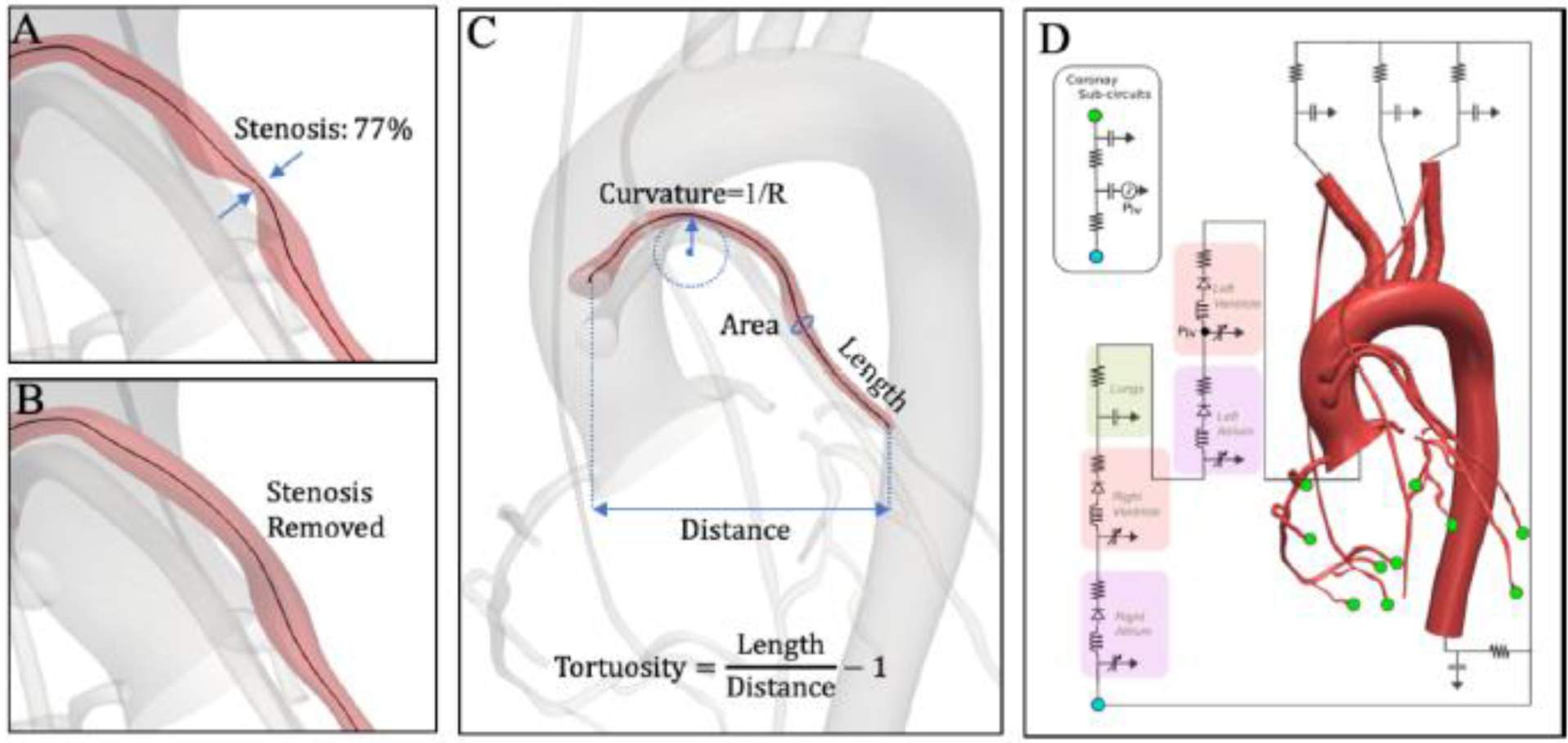
Figure A: Workflow showing CTA-based model reconstruction, stenosis removal, and extraction of anatomical and hemodynamic parameters for computational analysis.
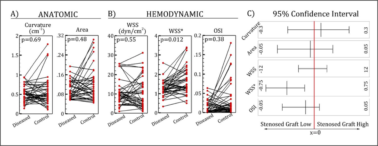
Figure B: Comparison of anatomical and hemodynamic parameters between diseased and control graft segments, including wall shear stress and oscillatory shear index.

The 3DQ Lab contributed by generating the anatomical models required for this analysis. Using post-surgical CTA data, vascular structures were segmented and reconstructed into 3D models using SimVascular. Approximately 75 patient-specific models were created, providing the foundation for CFD simulations and enabling detailed evaluation of graft hemodynamics across the study cohort.

These findings support the role of blood flow patterns in graft disease and highlight the potential of computational modeling to identify high-risk regions before clinical failure occurs. With further validation, this approach may help guide closer monitoring and more targeted treatment strategies for patients following bypass surgery.

Categories

  • Head & Neck
  • Chest
  • Abdomen & Pelvis
  • Upper Extremity
  • Lower Extremity
  • Case of the Month
  • Techniques

3DQ Lab – Grant Building

300 Pasteur Drive
Stanford, CA 94305

(650) 725-8432

Directions to Grant

3DQ Lab – Clark Building

318 Campus Drive
Stanford, CA 94305

(650) 725-6862

Directions to Clark

Learn More About the Lab

  • About Us
  • Meet the Team
Search Search

Copyright © Stanford University

© Copyright - 3D and Quantitative Imaging Laboratory - Enfold Theme by Kriesi
  • Link to LinkedIn
  • Link to Instagram
Link to: Early Detection of Transplant-Related Lung Injury with CT Link to: Early Detection of Transplant-Related Lung Injury with CT Early Detection of Transplant-Related Lung Injury with CT Link to: Fluorescence Tumor Imaging Link to: Fluorescence Tumor Imaging Fluorescence Tumor Imaging
Scroll to top Scroll to top Scroll to top