Get to Know the 3D Pro: Max Becket
Before joining the 3DQ Lab, Max Becket built a strong foundation in imaging and patient care. He grew up in a small rural town in south-central Pennsylvania and began studying radiologic technology soon after high school. While earning his associate’s degree, he worked weekends as a technologist assistant, gaining early experience in patient interaction and workflow. After graduating, he quickly became certified in CT and spent several years working at two Level 1 trauma and comprehensive stroke centers. By 2019, he had completed his bachelor’s degree in Health Sciences and was ready for a new challenge that combined his technical background with his interest in advanced visualization.
Max’s experience in CT gave him a deep appreciation for 3D visualization, and outside of work, he even took up 3D printing anatomical models as a hobby. When he began researching healthcare organizations that used this technology clinically, the 3DQ Lab quickly caught his attention. After following the lab’s work for a couple of years, an opening finally became available, and Max joined the team.
Right: Max Becket B.S. R.T.(R)(CT)
Today, Max is a 3D technologist specializing in structural heart imaging. He supports complex protocols such as TAVR, TMVR, TTVR, LAAO, and TRAC, where the smallest measurement can influence surgical decisions. These cases often involve quantifying valve dimensions, simulating implant placement, and visualizing how prosthetic devices will interact with surrounding anatomy.
He finds the work both challenging and deeply rewarding. Each study requires a detailed understanding of ECG-gated acquisitions, reconstruction techniques, and cardiac anatomy. As Max explains, processing the data is only part of the task; understanding what those measurements mean for the care team is what makes the work meaningful. To stay current, he regularly follows cardiac imaging lectures from the Society of Cardiovascular Computed Tomography and recommends Stanford Health Care’s YouTube tutorials on cardiac CT optimization for those looking to expand their skills.
Like many in the field, Max has seen a surge in imaging volume as technology becomes increasingly central to patient management. Staying efficient means continually refining workflows. The tools used in 3D imaging are advancing just as quickly as demand, and Max plays an active role in re-evaluating protocols to ensure every step adds value while maintaining quality.

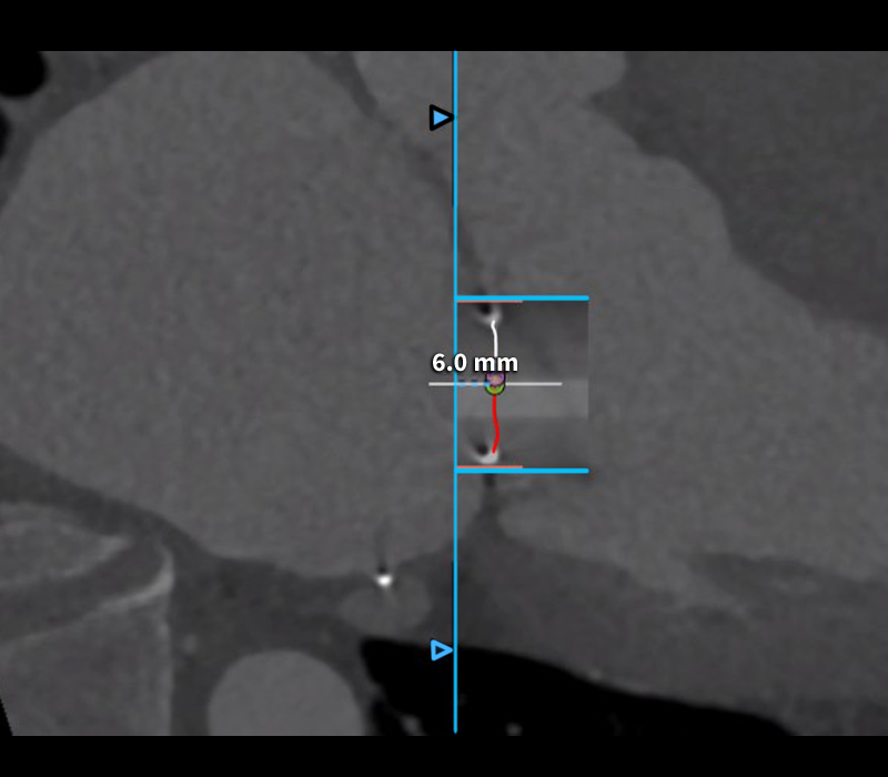
Figure B: Max is involved with creating valve measurements like these, which are provided to the TMVR team for evaluating prosthetic valve sizing and fitting.
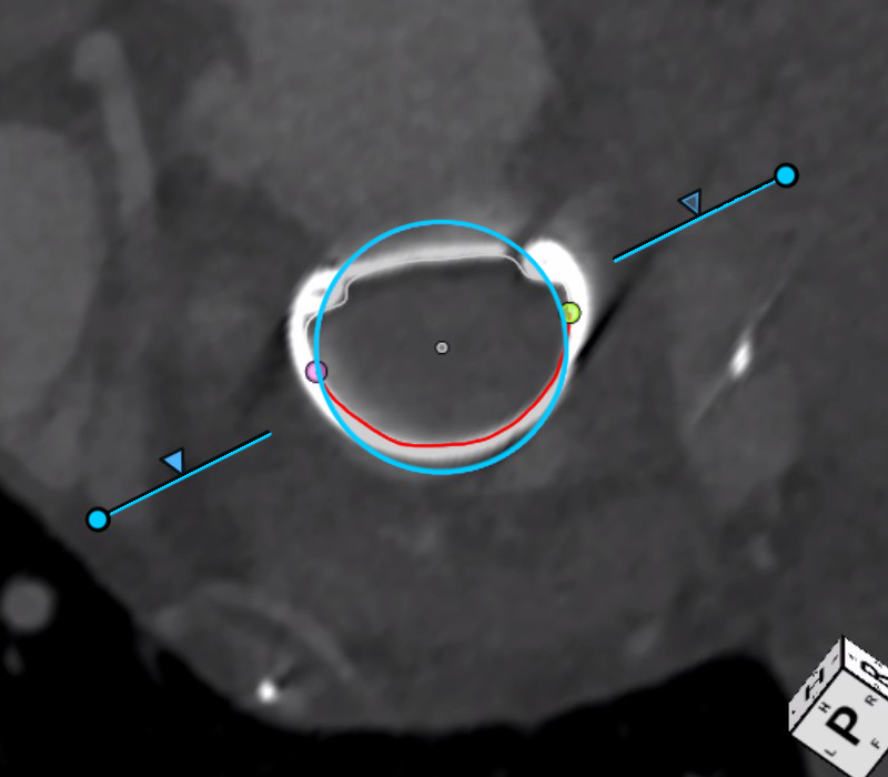
Figure C: Another example of TMVR measurements which are provided to the care team for evaluating prosthetic valve sizing and fitting.

