
Omental Flap Reconstruction
The omentum is a fatty apron-like structure within the abdomen that can be transferred as a vascularized flap for breast reconstruction. Stanford surgeons developed a novel technique that combines laparoscopic omental harvest with fat grafting to create a customized breast mound for patients who may not be candidates for more traditional reconstruction options.
In 2020, the Division of Plastic and Reconstructive Surgery approached the 3DQ Lab to explore whether CT imaging could be used to quantify breast volume before surgery. By measuring the breast with and without the overlying skin, the team could better estimate the amount of soft tissue required for reconstruction and compare that target volume with the tissue available from the omental flap.
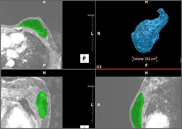
Figure A: Segmentation of the breast volume used for analysis, shown in cross-sectional CT images and segmentation (green) and as a volume render with calculated volume.
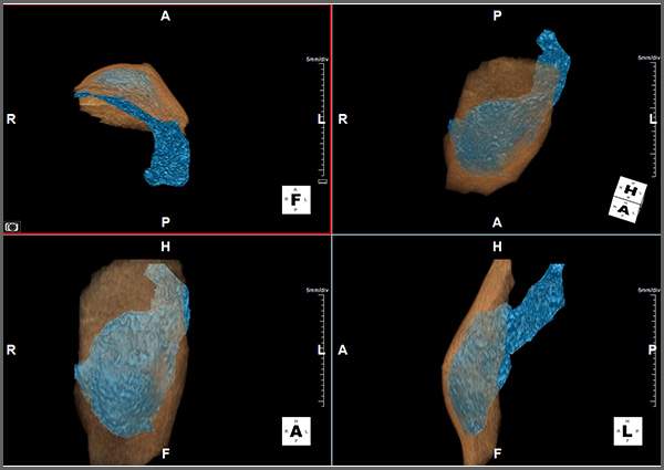
Figure B: Comparison of the segmented breast volume (blue) within the surrounding breast envelope (tan), used to estimate tissue requirements for omental flap reconstruction.