A patient was presented to the 3DQ Lab with right-sided hamstring pain that occurred only during intense activity, along with a drop in exercise ABIs. Because ABIs compare blood pressure at the ankle to the arm, a decrease seen only with exertion can suggest that blood flow becomes limited when the limb is in motion.
The patient also had a history of left iliac endofibrosis, a condition in which repetitive hip motion causes the arterial wall to thicken and the vessel to narrow. This had been treated with iliofemoral endarterectomy and angioplasty, procedures that remove diseased inner lining and widen narrowed segments.
Given the new exertional symptoms and concern for a motion-related arterial issue, additional imaging was requested of the 3DQ Lab for further evaluation and treatment planning.
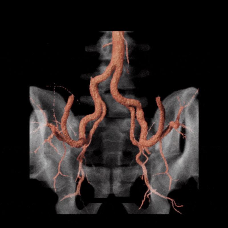
Figure A (Right): Rotational cinematic volume render of the iliac arteries (pink) and bone (transparent white) while in a stressed position.
A dynamic CTA was performed in both relaxed and stressed (flexed) positions to simulate the limb posture during activity. Changes in hip and pelvic motion can alter the course of the external iliac artery, and narrowing that affects blood flow may appear only when the limb is under tension. Imaging at rest alone may overlook these positional changes, so evaluating both postures helps determine whether the symptoms are related to dynamic vessel deformation rather than fixed structural disease. (Learn about a similar condition, popliteal entrapment, here).
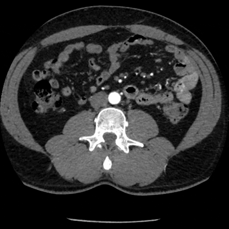
Figure B: Axial CT of the pelvis in a relaxed position.
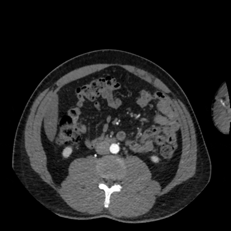
Figure C: Axial CT in a stressed position.
To better visualize the iliac anatomy, CPR loops were created to trace the artery along its course in both relaxed and stressed positions, allowing direct comparison of lumen size and curvature. VR views were generated with and without pelvic and femoral bone to show surrounding anatomical relationships. These complementary views help clarify how limb motion influences vessel shape and highlight narrowing that may be subtle or difficult to appreciate on axial slices alone. (Learn more about CPRs here, and VRs here).
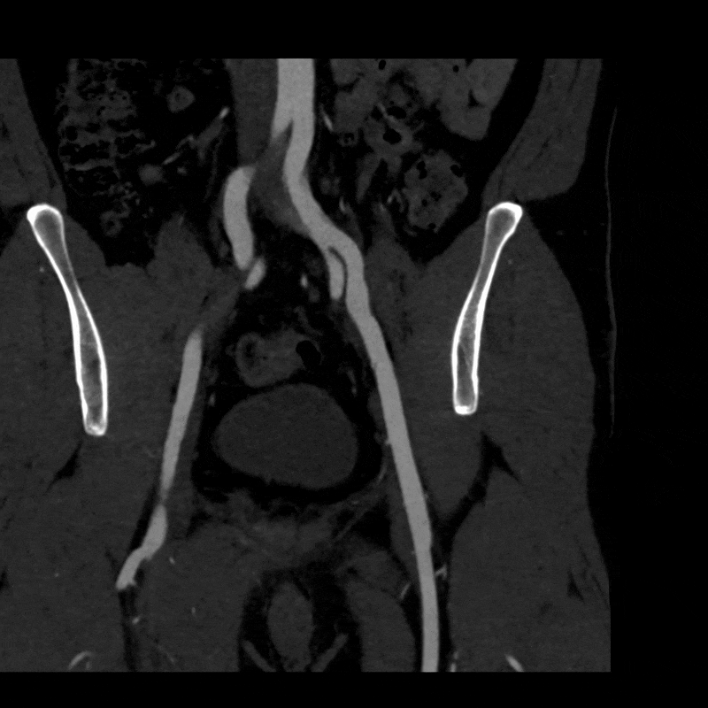
Figure D: CPR of the left iliac artery in a relaxed position.
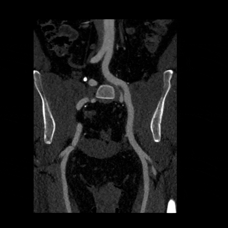
Figure E: CPR of the left iliac artery in a stressed position.
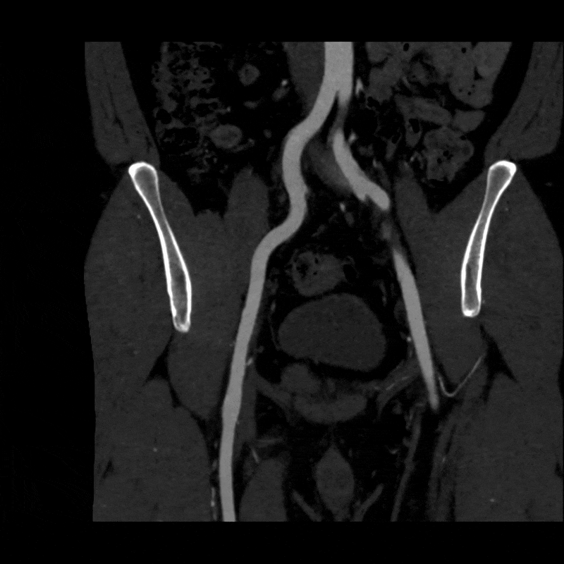
Figure F: CPR of the right iliac artery in a relaxed position.
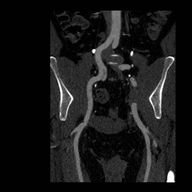
Figure G: CPR of the right iliac artery in a stressed position.
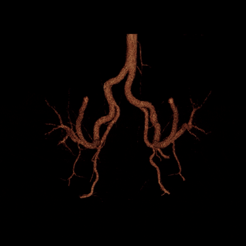
Figure H: Rotational volume render of the iliac arteries in the stressed position.
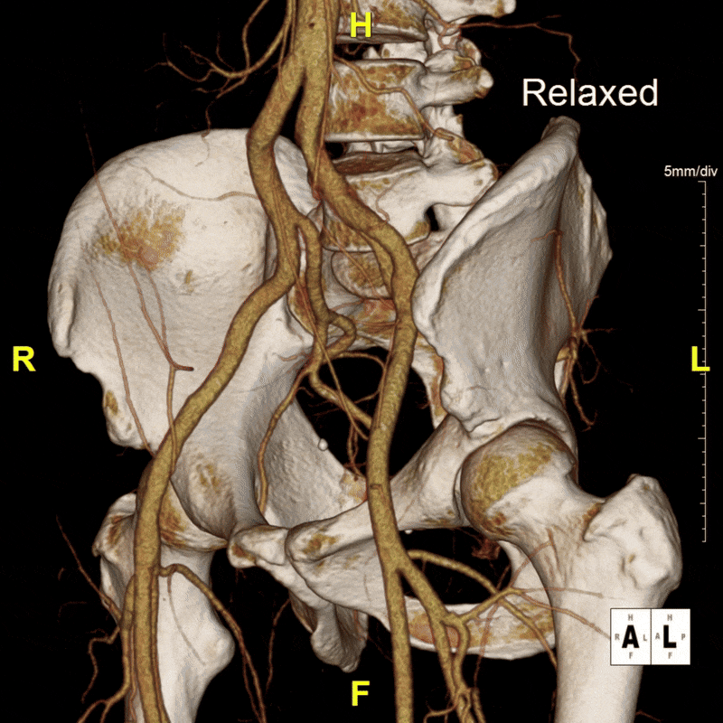
Figure I: Targeted volume renders of both the relaxed and stressed positions.
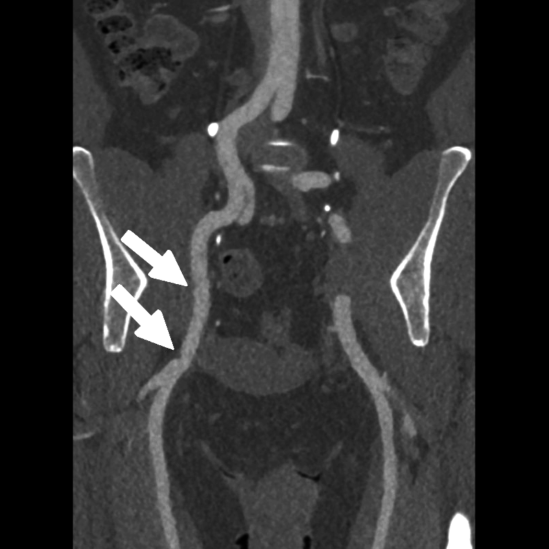
When the patient moved into the stressed position, the right external iliac artery showed two areas of luminal narrowing (a temporary decrease in the size of the vessel’s opening). One narrowing was more proximal (closer to the vessel’s origin near the pelvis) at about ten to twenty percent, and the second was more distal (farther along the vessel toward the leg) at about thirty to forty percent. Both returned to a widely patent appearance (fully open) when the leg moved back to a relaxed position.
The left external iliac artery remained patent across positions, and the common iliac arteries were tortuous (naturally curving) without any fixed stenosis. This pattern aligns with external iliac endofibrosis, where motion causes the artery to change shape rather than a fixed obstruction, highlighting why positional imaging can reveal abnormalities that may not appear on routine resting studies.
Figure J (Right): Two areas of narrowing were identified in the right external iliac.