
3D Printed Aortic Dissection Hemodynamic Models
Aortic dissection is a life-threatening condition where blood creates a tear in the aortic wall, entering and separating the layers of the aortic wall and creating a secondary channel known as the false lumen. Blood flow between the true and false lumens is heavily influenced by tears within the dissection flap, but the relationship between tear size, blood flow, and pressure changes remains difficult to evaluate in patients. Standard imaging can show anatomy, but understanding the complex hemodynamics that contribute to false lumen pressurization, vessel remodeling, and long-term risk remains challenging.
This study investigated how entry and exit tear size affect blood flow and pressure dynamics in a Type B aortic dissection. Researchers created models based on actual patient imaging and compared 4D-flow MRI measurements with fluid-structure interaction simulations to evaluate how changes in tear geometry altered true and false lumen flow patterns, pressure gradients, and vessel behavior. By combining controlled physical experiments with computational modeling, the study aimed to better understand the hemodynamic factors associated with disease progression and treatment plannin
Publication Link: Nature
Figure A: 3D rendering of the patient-specific aortic dissection model used for multi-material 3D printing and hemodynamic flow experiments.
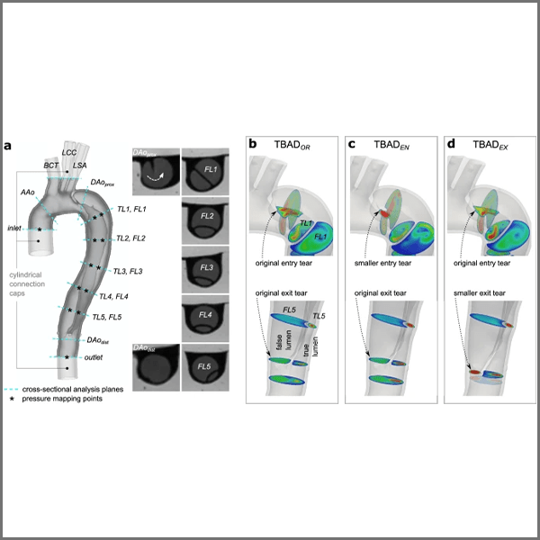
Figure B: 3D aortic dissection models showing tear modifications and analysis regions used for flow imaging and hemodynamic simulation experiments.
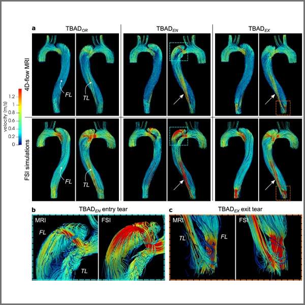
Figure C: 4D-flow MRI and simulation comparison showing blood flow patterns within the aortic dissection models under different tear configurations.