3D and Quantitative Imaging Laboratory
  • Home
  • Education
    • Education
    • Educational Post Feed
  • Research
    • Research
    • Validation Cohort: Acute Uncomplicated Type-B Aortic Dissection
  • Patient Care
    • Patient Care
    • 3D Printing in Medicine
    • 3DQ Lab TRAC Team (Tumor Response)
  • Case Studies
    • Case Studies
    • Innovation Projects
  • Click to open the search input field Click to open the search input field Search
  • Menu Menu

4D Flow MRI Pipeline for Pulmonary Hypertension

Collaborators: Stanford Engineering Marsden Lab

Chronic thromboembolic pulmonary hypertension (CTEPH) occurs when persistent blood clots obstruct the pulmonary arteries, increasing pressure in the lungs and placing strain on the right side of the heart. Although pulmonary endarterectomy can remove these obstructions, understanding how blood flow and vessel mechanics change after surgery remains challenging.

This study developed a semi-automated pipeline to analyze 4D flow MRI of the pulmonary arteries before and more than six months after pulmonary endarterectomy. The workflow quantified vessel size, wall stiffness, velocity, vorticity, and helicity to characterize how pulmonary artery flow patterns changed during recovery and how those changes related to improvements in pulmonary pressures and right ventricular function.

Publication Link: PubMed
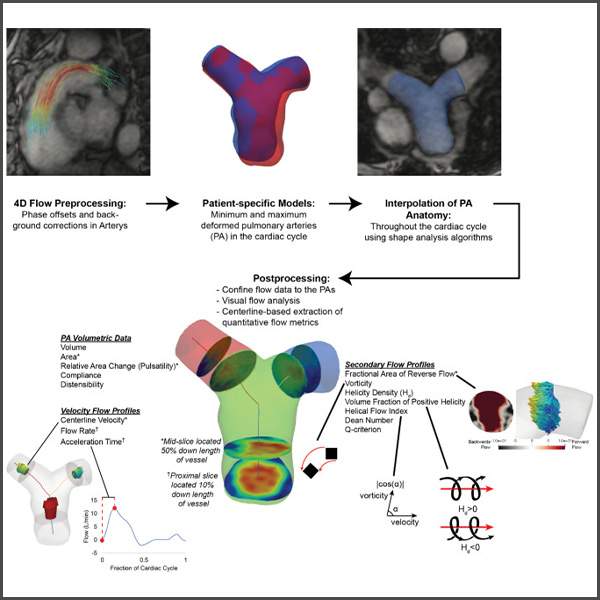
Figure A: Semi-automated 4D flow MRI analysis pipeline showing pulmonary artery segmentation, 3D model generation, and extraction of vessel stiffness and blood flow metrics.
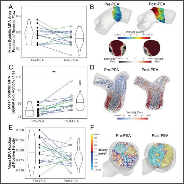
Figure B: Patient-specific pulmonary artery models from patients before and after pulmonary endarterectomy, illustrating the anatomical changes captured across more than 80 segmentations and 3D models created by the 3DQ Lab.

The 3DQ Lab created at least 80 patient-specific pulmonary artery segmentations and 3D models from 4D flow MRI, with additional models generated when needed to accurately track vessel motion throughout the cardiac cycle. These models formed the foundation of the post-processing pipeline used to extract blood flow and vessel stiffness metrics before and after surgery.

The study showed that successful surgery was associated with normalization of pulmonary artery size and stiffness, along with changes in complex flow patterns that correlated with right ventricular recovery. This work demonstrates how advanced imaging pipelines can provide noninvasive insight into cardiopulmonary remodeling and treatment response.

Categories

  • Head & Neck
  • Chest
  • Abdomen & Pelvis
  • Upper Extremity
  • Lower Extremity
  • Case of the Month
  • Techniques

3DQ Lab – Grant Building

300 Pasteur Drive
Stanford, CA 94305

(650) 725-8432

Directions to Grant

3DQ Lab – Clark Building

318 Campus Drive
Stanford, CA 94305

(650) 725-6862

Directions to Clark

Learn More About the Lab

  • About Us
  • Meet the Team
Search Search

Copyright © Stanford University

© Copyright - 3D and Quantitative Imaging Laboratory - Enfold Theme by Kriesi
  • Link to LinkedIn
  • Link to Instagram
Link to: 3D Printing for Pediatric Transplant Patients Link to: 3D Printing for Pediatric Transplant Patients 3D Printing for Pediatric Transplant Patients Link to: Silicone Heart Models for Septation Planning Link to: Silicone Heart Models for Septation Planning Silicone Heart Models for Septation Planning
Scroll to top Scroll to top Scroll to top