3D and Quantitative Imaging Laboratory
  • Home
  • Education
    • Education
    • Educational Post Feed
  • Research
    • Research
    • Validation Cohort: Acute Uncomplicated Type-B Aortic Dissection
  • Patient Care
    • Patient Care
    • 3D Printing in Medicine
    • 3DQ Lab TRAC Team (Tumor Response)
  • Case Studies
    • Case Studies
    • Innovation Projects
  • Click to open the search input field Click to open the search input field Search
  • Menu Menu

Siemens Auto Stitch & Batch
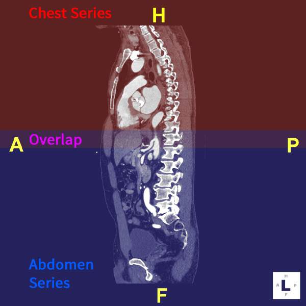
Some vendors’ CT protocols acquire the chest and abdomen as separate image series, even when they are intended to be interpreted as a single examination. For applications such as CT angiography, this can make it more difficult to review anatomy that spans both regions.

The 3DQ Lab explored whether these multipart studies could be automatically combined using TeraRecon’s Aquarius Processing Server (APS). Using DICOM tag filters, the system was configured to identify these specific examinations containing specific combinations of study descriptions and automatically generate a stitched dataset.

The goal was to create a single continuous image series that could then be batch processed and sent to PACS, allowing radiologists to review the combined dataset without requiring manual stitching by a technologist. This would have streamlined processing and improved access to whole-volume datasets for interpretation.

Although the project was not ultimately successful due to technical limitations, it represented an important attempt to automate a repetitive post-processing task. The work highlights the 3DQ Lab’s ongoing efforts to evaluate new approaches that can reduce manual effort and improve clinical workflows.

Learn more about stitching in our article here.

Figure A: Stitched CT dataset with color overlays indicating the portions derived from the chest (red) and abdominal (blue) series. The purple overlap region was used to align and merge the two acquisitions into a single continuous dataset.

Categories

  • Head & Neck
  • Chest
  • Abdomen & Pelvis
  • Upper Extremity
  • Lower Extremity
  • Case of the Month
  • Techniques

3DQ Lab – Grant Building

300 Pasteur Drive
Stanford, CA 94305

(650) 725-8432

Directions to Grant

3DQ Lab – Clark Building

318 Campus Drive
Stanford, CA 94305

(650) 725-6862

Directions to Clark

Learn More About the Lab

  • About Us
  • Meet the Team
Search Search

Copyright © Stanford University

© Copyright - 3D and Quantitative Imaging Laboratory - Enfold Theme by Kriesi
  • Link to LinkedIn
  • Link to Instagram
Link to: Non-Clinical Time Link to: Non-Clinical Time Non-Clinical Time Link to: Omental Flap Reconstruction Link to: Omental Flap Reconstruction Omental Flap Reconstruction
Scroll to top Scroll to top Scroll to top