
Common Measurement Techniques
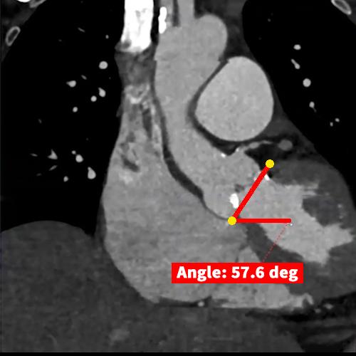
Angle: Angles are utilized in medical imaging to evaluate and measure the orientation, alignment, and relationships between anatomical structures within the human body. Angles are widely used in orthopedics, cardiology, vascular studies, and image registration.

Area: Area measurements involve quantifying the size of a two-dimensional surface or region. This is typically achieved by multiplying the length and width of each pixel within the selected region of interest and summing the values for all the pixels. In medical imaging, area measurements are frequently used to assess the cross-sectional area of vascular structures, particularly in conditions such as atherosclerosis, stenosis, or valvular disease.

Diameter: Diameter measurements represent a straight line usually passing through the center of a structure, representing its width. These measurements are vital for diagnosing conditions such as aneurysms, stenosis, or evaluating vascular or respiratory function.

Ejection Fraction: A percentage that represents the proportion of blood that is being pumped out of the heart with each heartbeat. Ejection fraction is calculated by dividing the volume of blood ejected from the left ventricle during systole (when the heart contracts) by the total volume of blood in the left ventricle at the end of diastole (when the heart relaxes).

Flow: Flow measurements are used to assess the direction, velocity, and volume of blood flow within blood vessels. Flow measurements are essential in evaluating cardiac function, assessing tissue perfusion, and quantifying blood flow rates in the aorta and major vessels.

Length: The distance between two specified endpoints. Length is commonly used in tumor and lesion analysis, vascular studies, skeletal measurements, anatomical dimensions, and surgical planning.

Perfusion: Quantification of the rate and distribution of blood flow in tissues or organs, providing valuable information about tissue viability, disease presence, and treatment response.

Perimeter: Perimeter measurements involve determining the length of a complex line drawn around a structure. Perimeter measurements are commonly utilized in medical applications such as lesion analysis, vascular studies, and valvular disease.
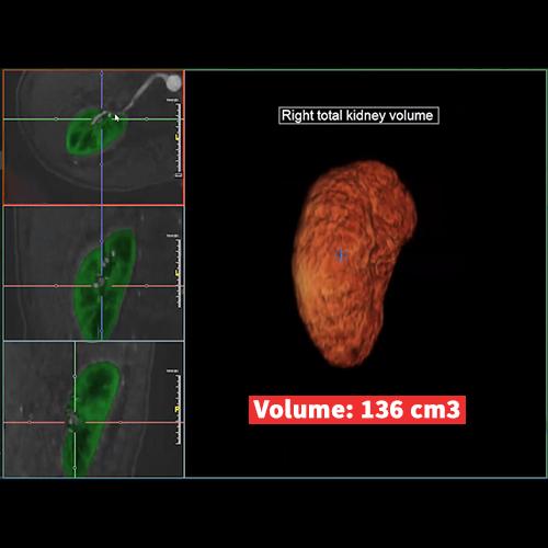
Volume: Volumetric measurements are essential for quantifying the size of three-dimensional structures. Medical professionals use this data to track changes in tumor growth, assess organ size for surgical planning, and more.

