
Allograft CT Scanning Workflow
Stanford’s musculoskeletal tumor service uses patient-specific surgical cutting guides to assist with complex bone tumor reconstruction procedures. These guides are designed from CT imaging and help surgeons plan how both the patient’s native bone and the donor bone, known as an allograft, should be cut to match during surgery.
Creating these guides requires high-resolution CT scans before surgery. This was initially difficult because the outside tissue bank supplying the allografts could not provide the necessary scans. To address this need, the Stanford 3DQ Lab worked with Stanford’s CT departments, physicist, and imaging teams to develop an internal allograft scanning protocol that continues to be used today.
Figure A: Axial CT images from a lower extremity allograft acquired using the Stanford 3DQ Lab allograft scanning workflow.
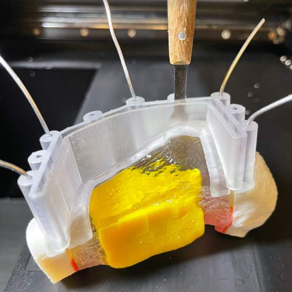
Figure B: Patient-specific surgical cutting guide positioned on a 3D printed allograft model to evaluate guide fit prior to surgery.