3D and Quantitative Imaging Laboratory
  • Home
  • Education
    • Education
    • Educational Post Feed
  • Research
    • Research
    • Validation Cohort: Acute Uncomplicated Type-B Aortic Dissection
  • Patient Care
    • Patient Care
    • 3D Printing in Medicine
    • 3DQ Lab TRAC Team (Tumor Response)
  • Case Studies
    • Case Studies
    • Innovation Projects
  • Click to open the search input field Click to open the search input field Search
  • Menu Menu

Vascular Access Training Models

Collaborators: Stanford Interventional Radiology

Ultrasound-guided vascular access procedures are performed by identifying a target vessel beneath the skin using an ultrasound probe, then advancing a needle or catheter into the vessel while monitoring placement in real time. Because arteries and deeper vessels can be difficult to access, procedural training on physical phantoms that simulate human anatomy can be a safe alternative to practicing on patients.

Many commercially available training phantoms can be expensive or provide limited ultrasound visibility. Interventional Radiology was interested in developing a more cost-effective model that could better reproduce vessel anatomy and echogenicity for procedural practice and education.
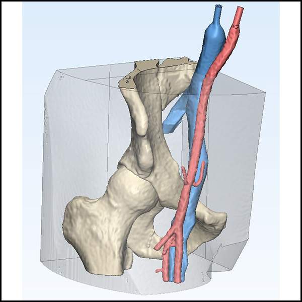
Figure A: Proposed vascular access phantom design showing iliac vasculature, pelvic anatomy, and the surrounding mold region used for ballistic gel casting.
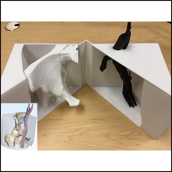
Figure B: Completed multi-material 3D printed phantom components designed to withstand the heat generated during ballistic gel casting.
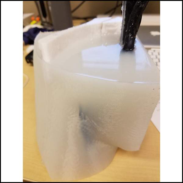
Figure C: Ballistic gel vascular access phantom with the flexible vascular model positioned inside the cast, prepared for ultrasound-guided procedural training.

The Stanford 3DQ Lab developed a custom vascular access phantom using segmented iliac vasculature, 3D printed structural components, and ballistic gel casting techniques. The iliac vessels were segmented from imaging data and printed in a flexible material using a Formlabs printer, while the surrounding mold and bony anatomy were designed by the lab and outsourced to the Rutt Lab for polycarbonate printing.

After fabrication, the printed components were provided to the Interventional Radiology team, who assembled the mold and performed the ballistic gel casting process. The reusable mold design allowed multiple training phantoms to be created by only replacing the vascular part and ballistic gel. The completed models were used for ultrasound-guided vascular access training and represent how imaging and additive manufacturing can support lower-cost procedural simulation and education.

Publication: ScienceDirect

Categories

  • Head & Neck
  • Chest
  • Abdomen & Pelvis
  • Upper Extremity
  • Lower Extremity
  • Case of the Month
  • Techniques

3DQ Lab – Grant Building

300 Pasteur Drive
Stanford, CA 94305

(650) 725-8432

Directions to Grant

3DQ Lab – Clark Building

318 Campus Drive
Stanford, CA 94305

(650) 725-6862

Directions to Clark

Learn More About the Lab

  • About Us
  • Meet the Team
Search Search

Copyright © Stanford University

© Copyright - 3D and Quantitative Imaging Laboratory - Enfold Theme by Kriesi
  • Link to LinkedIn
  • Link to Instagram
Link to: Allograft CT Scanning Workflow Link to: Allograft CT Scanning Workflow Allograft CT Scanning Workflow Link to: Nuss Procedure Training Model Link to: Nuss Procedure Training Model Nuss Procedure Training Model
Scroll to top Scroll to top Scroll to top