
Get to Know the 3D Pro: Derrick Laurel
Before joining the 3DQ Lab, Derrick Laurel earned a degree in psychology from San Francisco State and later trained as a radiologic technologist at Mills Peninsula Hospital. He worked in a wide range of roles—from sales and retail to research in a children’s mental health clinic—before deciding he wanted a more direct way to help people. Volunteering as a patient companion in Fremont confirmed that healthcare was the right path.
Derrick first encountered the 3DQ Lab during a tour in X-ray school, and when an entry-level position opened about a year later, he jumped at the chance. The learning curve was steep, but he was hooked—he even recalls dreaming about placing CPR points in vessels during those early weeks.
Right: Derrick Laurel, R.T. (R) | 3D Lab Technologist
Today, Derrick works across several advanced protocols including neurosurgical guidance, fMRI, cardiovascular studies, and airway cases. He also leads efforts around integrating AI into radiology workflows, helping assess and implement new tools.
One of Derrick’s favorite protocols is the lower extremity runoff—a CT angiogram that captures arterial flow from the abdomen to the feet to evaluate peripheral artery disease. It’s a technically demanding study, and Derrick compares the post-processing to a video game: carefully placing curved planar reformation (CPR) paths and using the “circling” technique to segment vessels from surrounding bone. Processed runoff images such as MIPs or CPRs may allow clinicians to view the full length of a vessel in a single image and quickly identify areas of narrowing or occlusion (seen below).
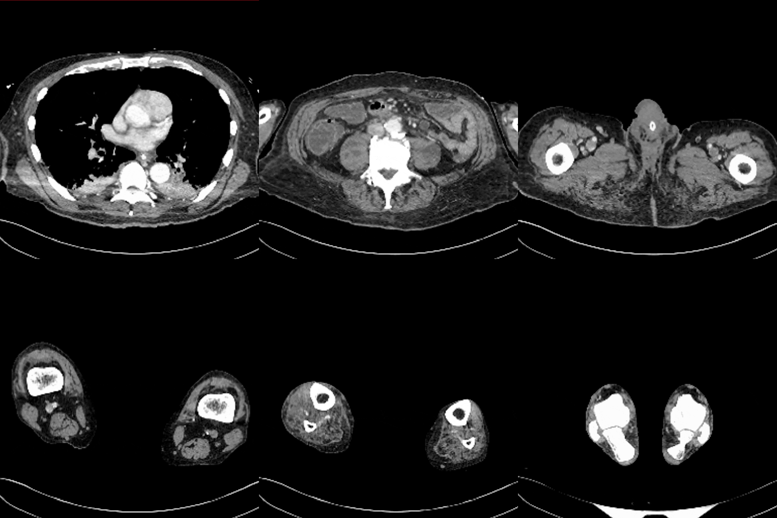
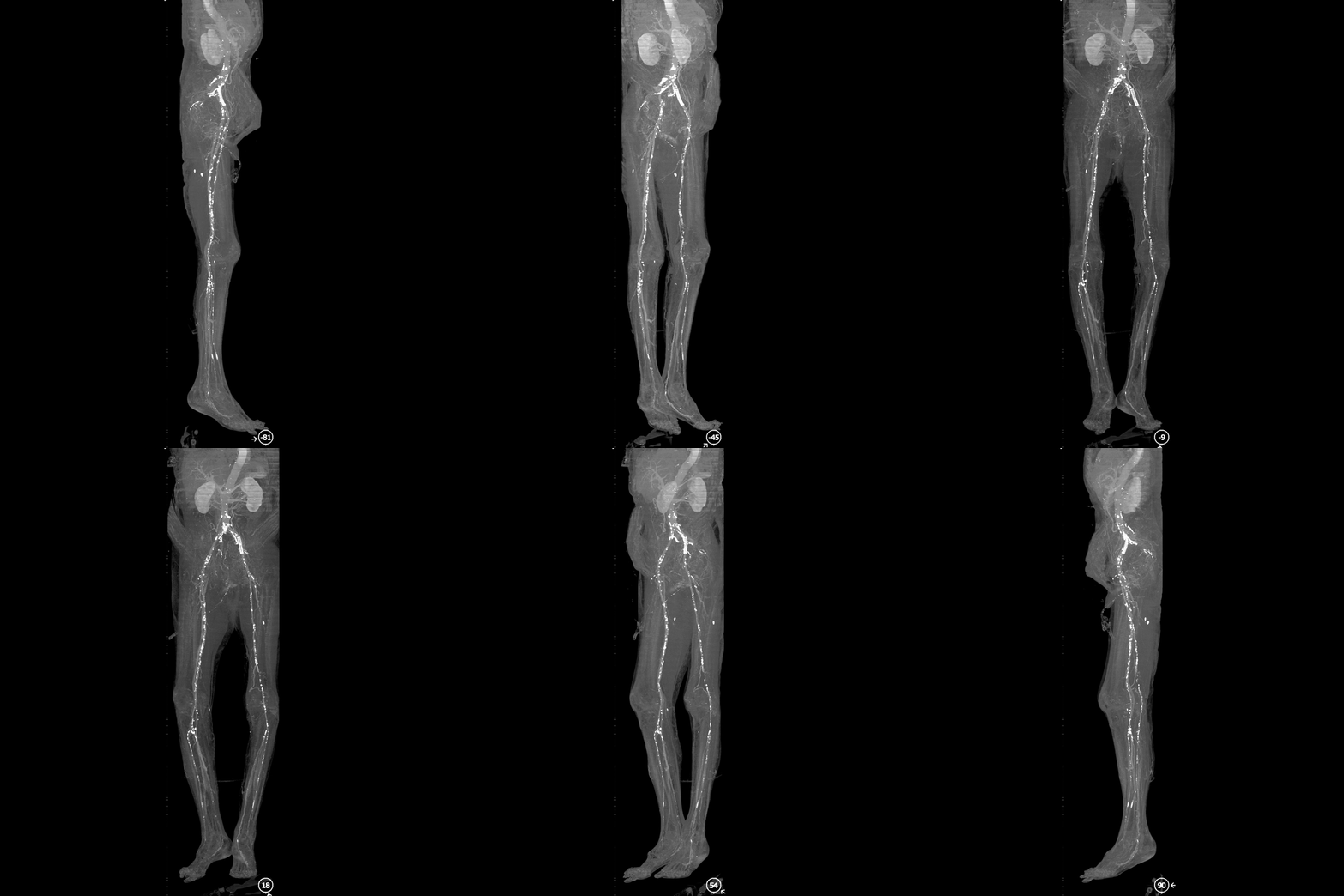
Figure B: MIP images highlight the brightest voxels along the viewing axis, helping visualize vessel narrowing and calcified plaque across long arterial segments in this lower extremity runoff.
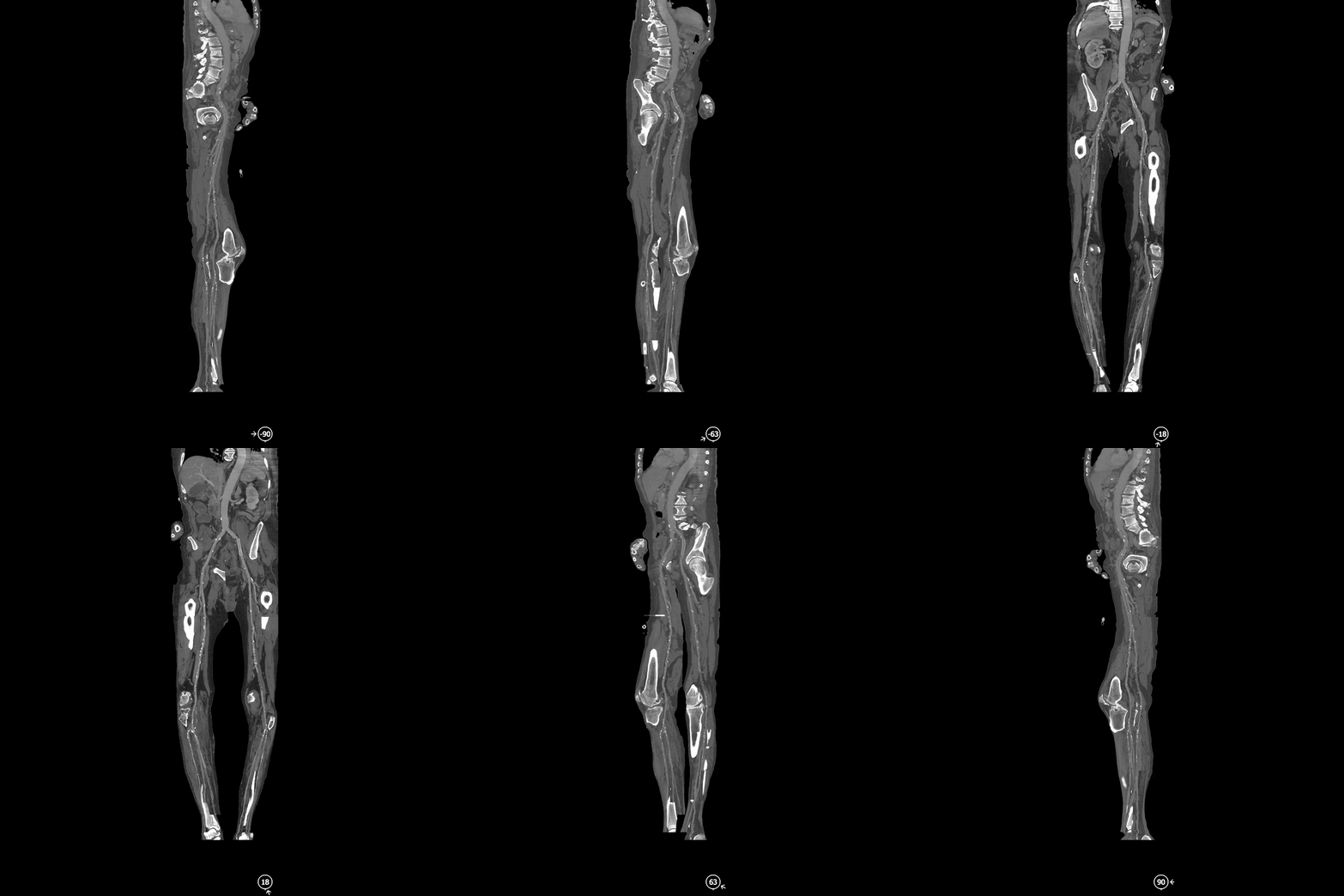
Figure C: Multipath CPR image of the lower extremities. These reconstructions are often generated from runoff datasets to visualize each vessel individually and allow for quick evaluation of occlusions or narrowing.

