3D and Quantitative Imaging Laboratory
  • Home
  • Education
    • Education
    • Educational Post Feed
  • Research
    • Research
    • Validation Cohort: Acute Uncomplicated Type-B Aortic Dissection
  • Patient Care
    • Patient Care
    • 3D Printing in Medicine
    • 3DQ Lab TRAC Team (Tumor Response)
  • Case Studies
    • Case Studies
  • Search
  • Menu Menu

What Is Dual Energy CT?

CT is a powerful tool for capturing anatomy, but it has limits. The limits are especially apparent when different tissues appear similar on scans or when metal implants create artifacts. In these cases, it can be difficult to determine what’s actually happening inside the body. Inflammation, infection, and tumors may appear nearly identical on standard CT, and subtle differences in tissue composition can be missed. This can lead to additional imaging or even invasive procedures to reach a diagnosis.

Dual energy CT (DECT) helps overcome these challenges. Unlike traditional CT, which uses a single X-ray energy level, dual energy CT captures images at two. This is important because different materials absorb low- and high-energy X-rays in distinct ways. The resulting datasets are processed using specialized algorithms that compare how tissues respond across both energy levels, allowing for more accurate differentiation. This enables visualization of iodine uptake in tumors, clearer distinction between tissue types, and reduction of metal artifacts that would otherwise obscure important details.

Applications

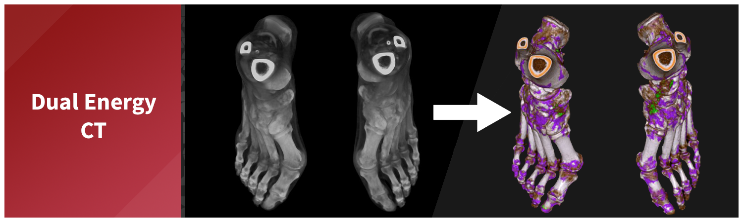
Dual energy CT has a wide range of clinical applications, and our lab uses it regularly to enhance diagnostic clarity across multiple areas. Two common examples include characterizing kidney stones and identifying urate crystal deposits in cases of suspected gout, both of which benefit from dual energy’s ability to distinguish materials that appear similar on standard CT.

Gout

Gout is a type of inflammatory arthritis caused by a buildup of uric acid crystals in the joints and surrounding tissues. These deposits trigger painful flares and long-term joint damage if left untreated. One of the key challenges in diagnosing gout is distinguishing uric acid crystals from other materials—particularly calcium deposits, which can appear similar on standard CT scans. This distinction is critical, as treatments for gout differ significantly from those used for calcium-based conditions like pseudogout or calcific tendinitis. Without a clear diagnosis, patients may receive the wrong treatment or face delays in care.

Dual energy CT addresses this problem by capturing images at two different X-ray energy levels. Because uric acid and calcium absorb X-rays differently depending on the energy, DECT can separate the two and highlight urate deposits directly on the images. This gives clinicians a non-invasive way to confirm gout, especially in difficult cases where fluid aspiration isn’t possible. DECT can also measure the total volume of crystal burden, allowing physicians to track how well urate-lowering therapies work over time. By improving both diagnosis and disease monitoring, DECT supports more accurate, tailored treatment for patients with gout.

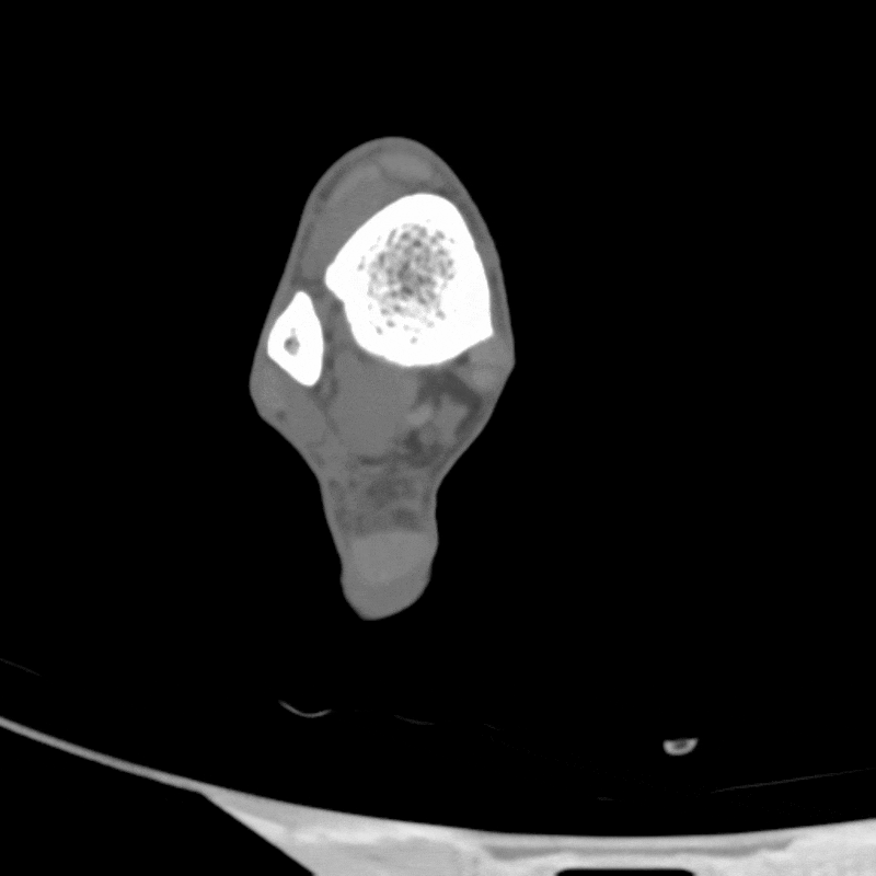
Figure A: Dual energy CT images of the foot, to be used for identifying gout.

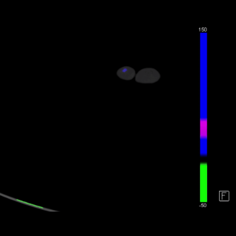
Figure B: The images of the foot from Figure A after post-processing filters have been applied, highlighting the presence of urate crystals in purple and calcified structures in green.

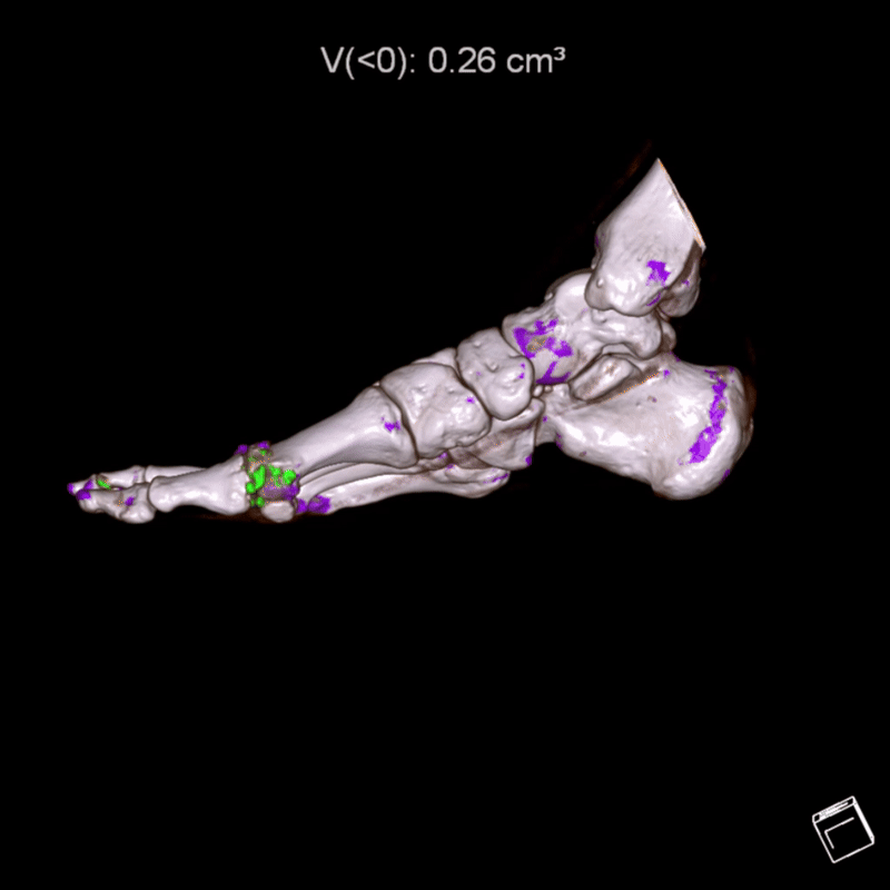
Figure C: Volume rendering of the imaging from Figure B, demonstrating the extent of urate crystal and calcified structures in 3D.

Kidney Stones

Kidney stones are hard mineral deposits that form in the kidneys and can cause severe pain, bleeding, or infection when they obstruct the urinary tract. Treatment depends heavily on the type of stone, as different compositions respond to different therapies. For example, uric acid stones can often be dissolved with medication, while calcium-based stones typically require physical removal. Standard CT is excellent at detecting stones, but it cannot reliably tell one type from another, leaving clinicians to rely on guesswork or post-removal analysis.

Dual energy CT offers a major improvement by allowing radiologists to identify stone composition non-invasively. By capturing images at two X-ray energy levels, it distinguishes uric acid from calcium and other stone types based on their material-specific attenuation patterns. This helps guide treatment from the start, determining whether a patient may benefit from medical management or needs surgical intervention. It can also reduce the need for repeat imaging or exploratory procedures. For patients with recurrent stones, DECT provides valuable insight into stone behavior over time, supporting more personalized prevention strategies.

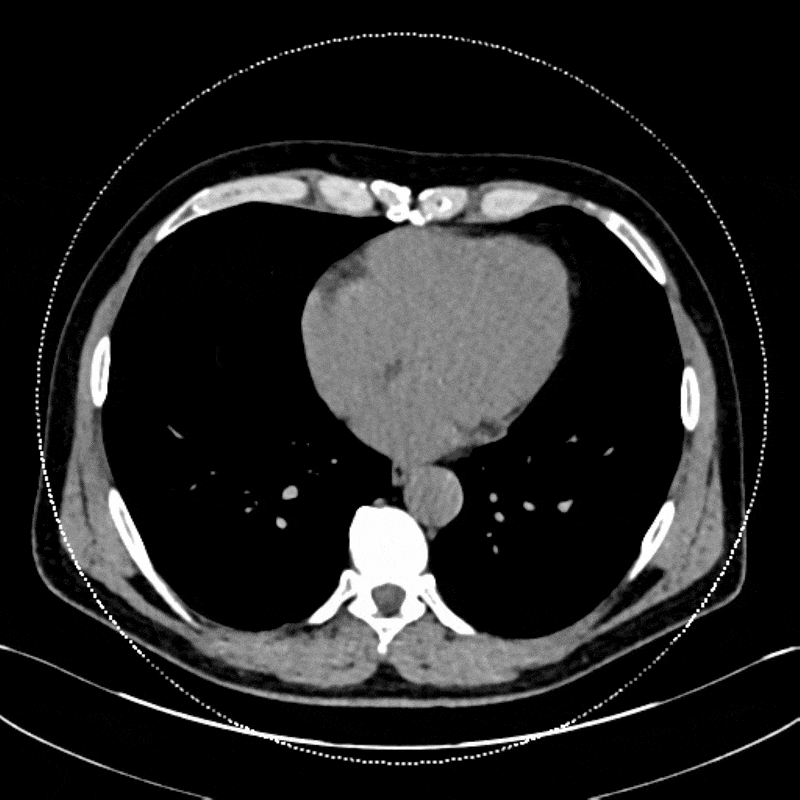
Figure D: Axial dual energy CT images of the abdomen, used for analyzing the composition of kidney stones.

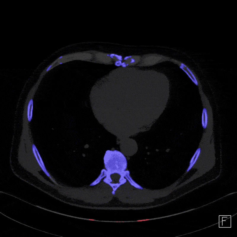
Figure E: The axial images of the abdomen from Figure D, now processed to analyze the composition of the kidney stone (indicated by the arrow).

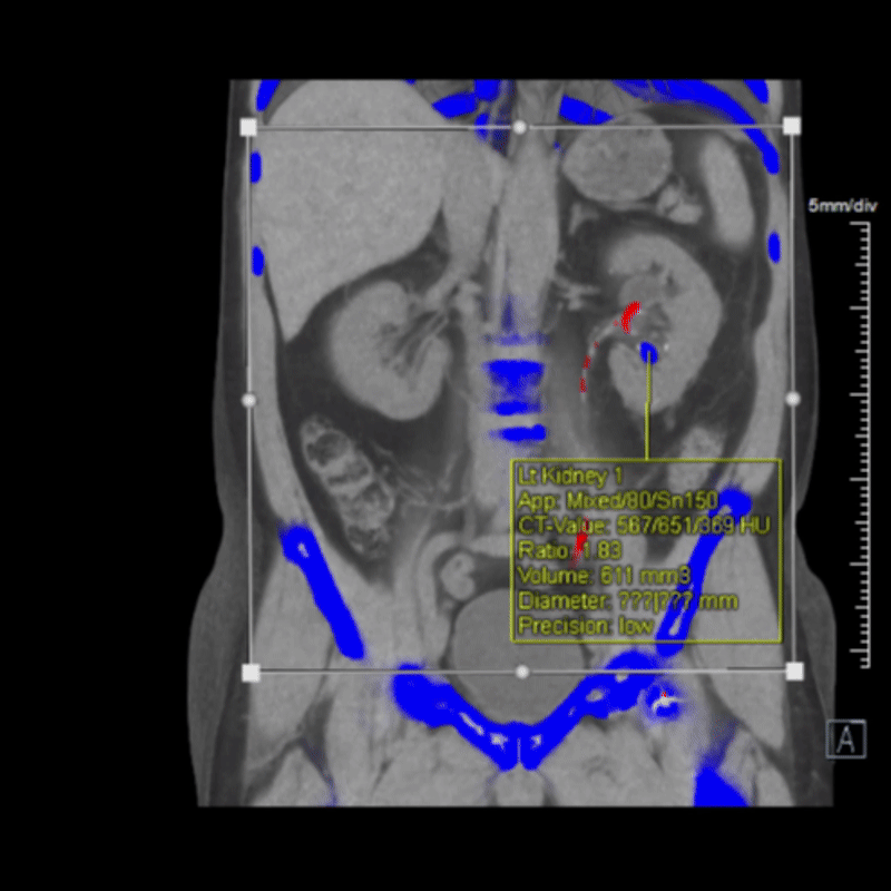
Figure F: Processed images from Figure E, showing graphics that detail the stone’s size, volume, and composition.

Challenges of CT Dual Energy

Limitations  
While dual energy CT offers major advantages, it does have some trade-offs. Image noise can be higher, especially at lower energy levels, which may reduce clarity in certain patients, particularly those with larger body types. Not all tissues or conditions benefit equally from dual energy separation, so in some cases, it doesn’t add much beyond standard CT.

Radiation Exposure
Early dual energy CT systems did increase radiation exposure compared to standard CT because they often performed two scans or used less efficient detectors. Fortunately, modern dual energy systems are designed to keep radiation doses similar to, or even lower than a regular CT. Many now use dual-layer detectors or rapid kVp switching to collect both datasets at once without needing extra exposure. So, if performed on newer machines, radiation is generally not a major concern.

Cost
Yes, dual energy CT is more expensive. The scanners themselves cost more, the software and post-processing tools add cost, and interpreting dual energy studies often takes additional radiologist time. That said, in some cases, it can replace multiple imaging studies, which may save costs overall, but the upfront expense may be higher.

Future of CT Dual Energy

The role of dual energy CT in clinical imaging continues to grow, with expanding applications and increasing integration into routine protocols. As the technology matures, its ability to improve tissue characterization, reduce artifacts, and guide more precise treatment decisions is becoming widely recognized across multiple specialties. Advances in automation, including machine learning tools for material classification and interpretation, are also helping streamline its use in practice.

Looking ahead, new detector technologies like photon-counting CT are poised to carry these capabilities even further. By capturing energy data at the level of individual photons, photon-counting systems offer greater precision, improved image quality, and potential for lower radiation dose. These next-generation systems build on the foundation of dual energy CT and may help expand its role even more in the years to come.

Categories

  • Head & Neck
  • Chest
  • Abdomen & Pelvis
  • Upper Extremity
  • Lower Extremity
  • Case of the Month
  • Techniques

3DQ Lab – Grant Building

300 Pasteur Drive
Stanford, CA 94305

(650) 725-8432

Directions to Grant

3DQ Lab – Clark Building

318 Campus Drive
Stanford, CA 94305

(650) 725-6862

Directions to Clark

Learn More About the Lab

  • About Us
  • Meet the Team

Copyright © Stanford University

© Copyright - 3D and Quantitative Imaging Laboratory - Enfold Theme by Kriesi
  • LinkedIn
  • Instagram
Get to Know the 3D Pro: Derrick LaurelDIEP (Deep Inferior Epigastric Perforator) Flap Reconstruction
Scroll to top