What Are Metal Artifacts and Why Do They Matter?
CT imaging is a widely used diagnostic tool in modern medicine, enabling detailed visualization of the body’s internal structures. However, the presence of metal implants often complicates the diagnostic viability of CT scans due to the physics of CT. Interaction between X-rays and metallic objects within the body, such as joint replacements, dental fillings, or surgical screws and plates can lead to visual anomalies called metal artifacts. This occurs because metallic objects have a higher density compared to human tissues, causing them to absorb and scatter X-rays more intensely. This interaction leads to various imaging distortions, including streaking, beam hardening, and signal loss, which can degrade the quality of the CT images.
Streaking appears as lines radiating from the metal object, while beam hardening manifests as dark bands or areas that look falsely more dense than they actually are. These phenomena can obscure critical anatomical details near the metal, complicating the diagnosis of conditions and the assessment of post-surgical recovery. For instance, in orthopedic imaging, metal artifacts can hide signs of infection or loosening of prosthetic joints, whereas in dental scans, they can mask underlying bony pathology.
Figure A: Axial CT imaging of a knee with a metallic implant, demonstrating metal artifact.
How Do We Reduce Metal Artifacts?
Several strategies have been developed to mitigate metal artifacts in CT imaging, enhancing both the clarity and utility of the scans:
• Metal Artifact Reduction Software (MAR): This software specifically targets and corrects the distortions caused by metal objects in CT scans. It adjusts for altered X-ray paths that occur around metal implants, thereby improving the clarity of the surrounding tissue in the images.
• Dual-Energy CT (DECT): This technique uses two different X-ray energy levels during the scan. The varying absorption rates of metal and soft tissues at these energies allow the system to differentiate between these materials more effectively, which helps in reducing artifacts and enhancing image clarity.
• Iterative Reconstruction: This is an advanced algorithmic approach that reconstructs the imaging data multiple times to improve accuracy. Each iteration refines the image by reducing noise and compensating for missing or distorted data, including those affected by metal objects.
While these techniques collectively represent advancements in the management of metal artifacts, they do not completely eliminate them. Residual artifacts can still occur, particularly in cases involving large, dense, or complex metallic implants. These remaining artifacts may still obscure critical details and pose challenges in accurately interpreting CT scans.

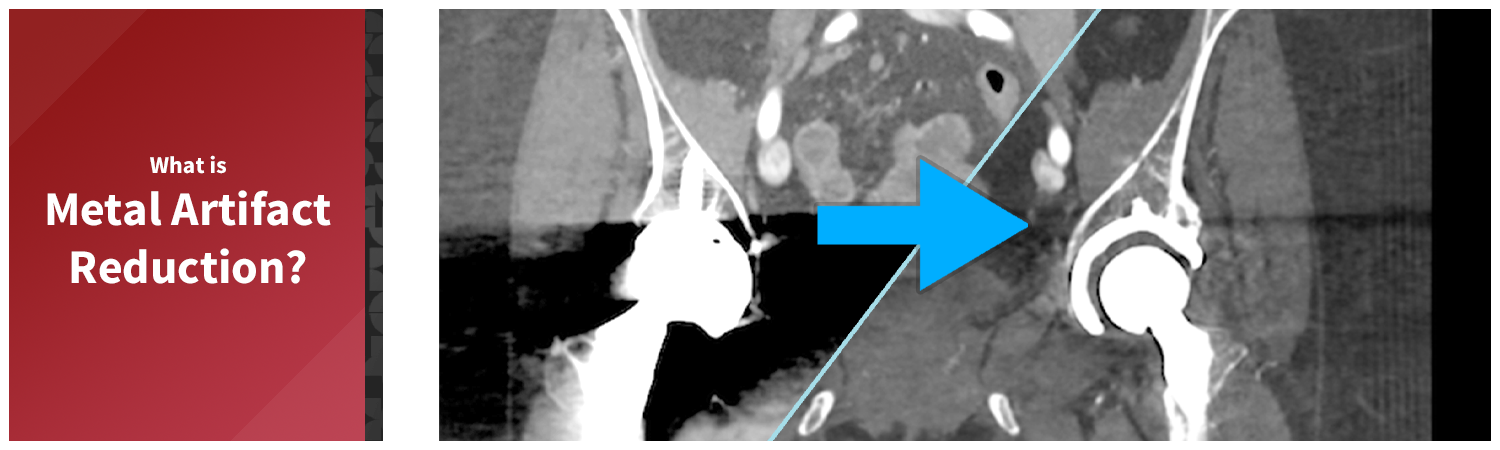
Figure B: Coronal view of a CT scan with significant metal artifact distortion around the pelvis due to hip replacement hardware.
Figure C: The same coronal imaging from figure B, after metal artifact reduction algorithms have been applied.
Affect of Metal Artifacts in 3D Imaging
Metal artifacts can significantly hinder both the accuracy of measurements and the clarity of volume renders created for 3D imaging. The streaks and shadows caused by metal artifacts within the scan field can alter the perceived dimensions and relationships of anatomical structures. This distortion can lead to inaccuracies in measuring critical dimensions, such as the size and position of tumors or the alignment of prosthetic implants. Furthermore, metal artifacts can degrade the overall quality of 3D visualizations, making them lose their ability to be tool for quickly assessing a targeted anatomical region. Metal artifact-reduced images are preferred for 3D imaging because they provide clearer and more accurate visualizations of anatomical structures.
Figure D (Right): Targeted views and measurements created for Transcatheter Heart Valve (THV) evaluation on scans both with and without metal artifact reduction (Figures B & C). The effects of metal artifacts can be seen in the difference of image quality as well as anatomical size and positioning.
Figure E: Volume rendering of the imaging from Figure B, demonstrating the effect of metal artifacts on the visual fidelity of 3D imaging.
Figure F: Volume rendering of the imaging from Figure C, showcasing the improvement after applying metal artifact reduction. This provides a clear visual contrast to Figure D, which displays the original 3D imaging without metal artifact reduction.

