3D and Quantitative Imaging Laboratory
  • Home
  • Education
    • Education
    • Educational Post Feed
  • Research
    • Research
    • Validation Cohort: Acute Uncomplicated Type-B Aortic Dissection
  • Patient Care
    • Patient Care
    • 3D Printing in Medicine
    • 3DQ Lab TRAC Team (Tumor Response)
  • Case Studies
    • Case Studies
    • Innovation Projects
  • Click to open the search input field Click to open the search input field Search
  • Menu Menu

TAVR Meeting

Collaborators: Stanford TAVR Program

Patients being evaluated for Transcatheter Aortic Valve Replacement (TAVR) are often reviewed by a large multidisciplinary team that includes surgeons, interventionalists, imaging specialists, nursing staff, coordinators, and other clinical services. Because procedural planning depends on both anatomical imaging and patient-specific clinical factors, there is a need for a collaborative review process that allows all teams to evaluate cases together efficiently and align on treatment decisions.

The Stanford 3DQ Lab generates the post-processed images and measurements used for TAVR evaluation, leading 3DQ technologists to become actively involved in these planning meetings. Meetings were originally held within the 3DQ Lab in an office space tailored for multidisciplinary case review, with multiple displays used simultaneously for live 3D imaging, echocardiography review, measurements, and patient clinical information. Additional seating and workspace accommodations were arranged to support participation from the entire clinical team.

Figure A. Video clip demonstrating TAVR measurements and images created by the Stanford 3DQ Lab for procedural planning.
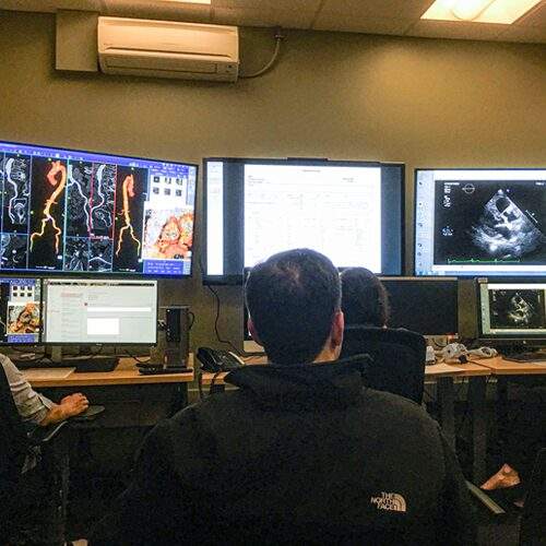
Figure B. In-person  TAVR planning meeting held within the Stanford 3DQ Lab office.

During the meeting, clinicians frequently request additional anatomical views or measurements to evaluate vessel tortuosity, narrowing, calcification, access feasibility, and valve sizing considerations in real time. The meetings are highly interactive, with surgeons, interventionalists, nursing teams, patient coordinators, and specialists collaborating on procedural planning.

Following the COVID-19 pandemic, the workflow transitioned to a fully virtual format while continuing to support multidisciplinary case review. The meetings remain an example of how advanced imaging, quantitative analysis, and collaborative review are integrated into structural heart procedural planning.

Learn more about TAVR in our article here.

Categories

  • Head & Neck
  • Chest
  • Abdomen & Pelvis
  • Upper Extremity
  • Lower Extremity
  • Case of the Month
  • Techniques

3DQ Lab – Grant Building

300 Pasteur Drive
Stanford, CA 94305

(650) 725-8432

Directions to Grant

3DQ Lab – Clark Building

318 Campus Drive
Stanford, CA 94305

(650) 725-6862

Directions to Clark

Learn More About the Lab

  • About Us
  • Meet the Team
Search Search

Copyright © Stanford University

© Copyright - 3D and Quantitative Imaging Laboratory - Enfold Theme by Kriesi
  • Link to LinkedIn
  • Link to Instagram
Link to: Nuss Procedure Training Model Link to: Nuss Procedure Training Model Nuss Procedure Training Model Link to: Protocol Management Link to: Protocol Management Protocol Management
Scroll to top Scroll to top Scroll to top