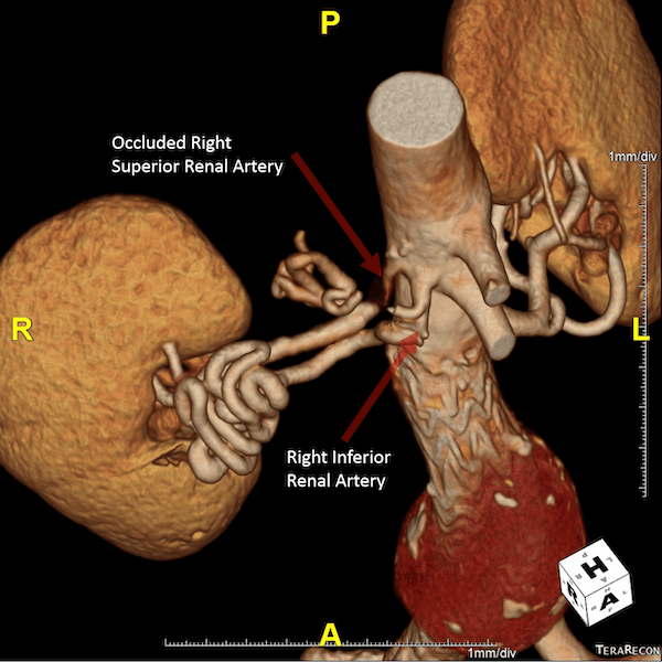
68 year old male with a history of a stented abdominal aortic aneurysm. CT scan with IV contrast reveals the stent graft placement and aneurysm thrombus. There was also an incidental finding of extensively convoluted right inferior renal artery due to a young-age occlusion of the right superior renal artery. These 3D volume rendered models produced by one of our technologists clearly demonstrates these vascular abnormalities for the patient and his doctors. Production of the images and video below requires advanced software and our lab’s skilled technologists.


Rossi Patrick
3DQ Technologist