3D and Quantitative Imaging Laboratory
  • Home
  • Education
    • Education
    • Educational Post Feed
  • Research
    • Research
    • Validation Cohort: Acute Uncomplicated Type-B Aortic Dissection
  • Patient Care
    • Patient Care
    • 3D Printing in Medicine
    • 3DQ Lab TRAC Team (Tumor Response)
  • Case Studies
    • Case Studies
    • Innovation Projects
  • Click to open the search input field Click to open the search input field Search
  • Menu Menu

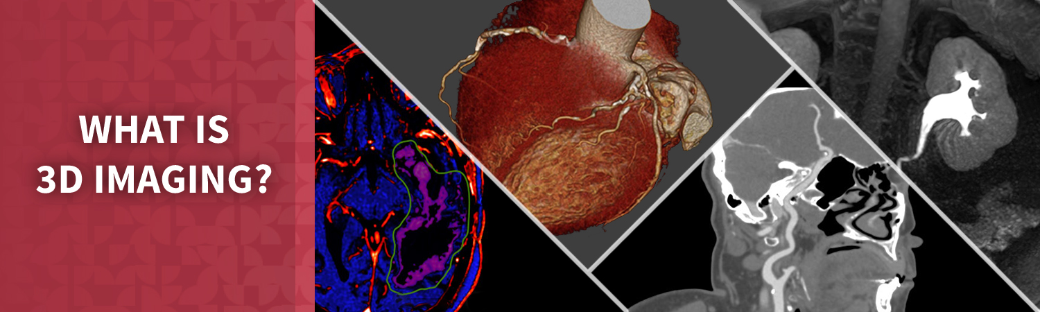
What is 3D Imaging?

3D Imaging in healthcare is a collection of techniques and tools used to post-process medical scan data. Unlike 2D imaging, which offers flat, linear views limited to length and width, 3D imaging adds depth, providing a comprehensive representation in three dimensions: length, width, and height (x, y, z coordinates). This depth enables detailed anatomical visualizations of the human body, using technologies such as CT, MRI, and Ultrasound. While 2D methods like X-rays and standard ultrasounds are effective for general imaging, they lack the capability to offer the thorough perspective that 3D imaging provides. Since its introduction in the 1990s it has revolutionized medical diagnosis and treatment, giving healthcare professionals deeper insights into complex health conditions.

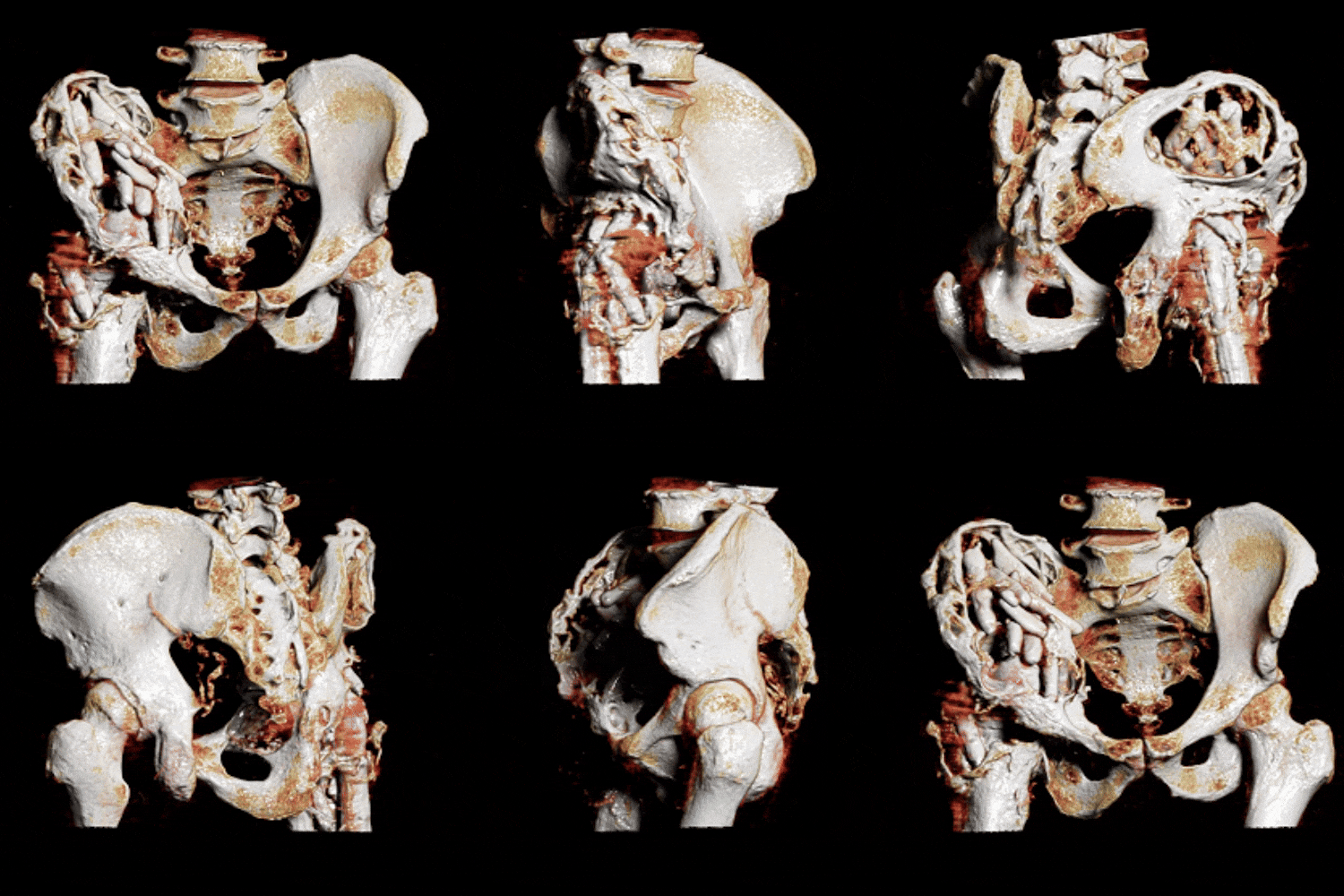
Figure A (Right): 3D volume rendering of a degenerative pelvis created from CT scan data.
How is 3D Imaging Created?

The creation of 3D imaging in healthcare is a sophisticated process that hinges on several elements. It begins with scanning a patient’s anatomy based on clinical concern. The quality of this scan data is crucial; it follows the “garbage in, garbage out” principle, meaning that the accuracy and resolution of the initial scan data directly impacts the quality of the 3D imaging (1)(2). Key elements like high resolution, contrast enhancement, and the minimization of artifacts significantly influence the overall quality of both the imaging scan and 3D. These aspects should be evaluated before performing the scan to ensure the resulting 3D image is as precise and informative as possible. 

The completed scan is loaded into FDA-approved medical imaging software by a radiologist or radiologic technologist, who can use it to produce a variety of 2D and 3D images. This software generally requires at minimum some manual guidance, and it’s becoming more common to see automations such as artificial intelligence to assist with reconstruction. The role of radiologists is crucial as they not only oversee the imaging process but also interpret the results. 

Below are some examples of the way the 3DQ Lab uses 3D Imaging to enhance patient care; each image will link you to a page related to the topic:

Image Processing

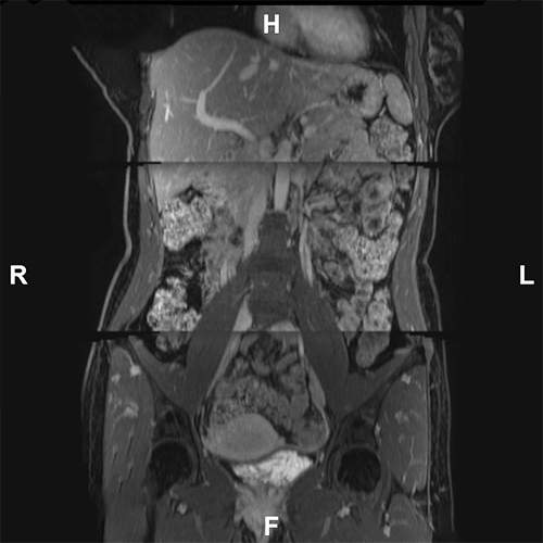
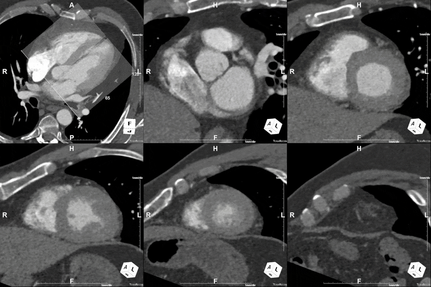
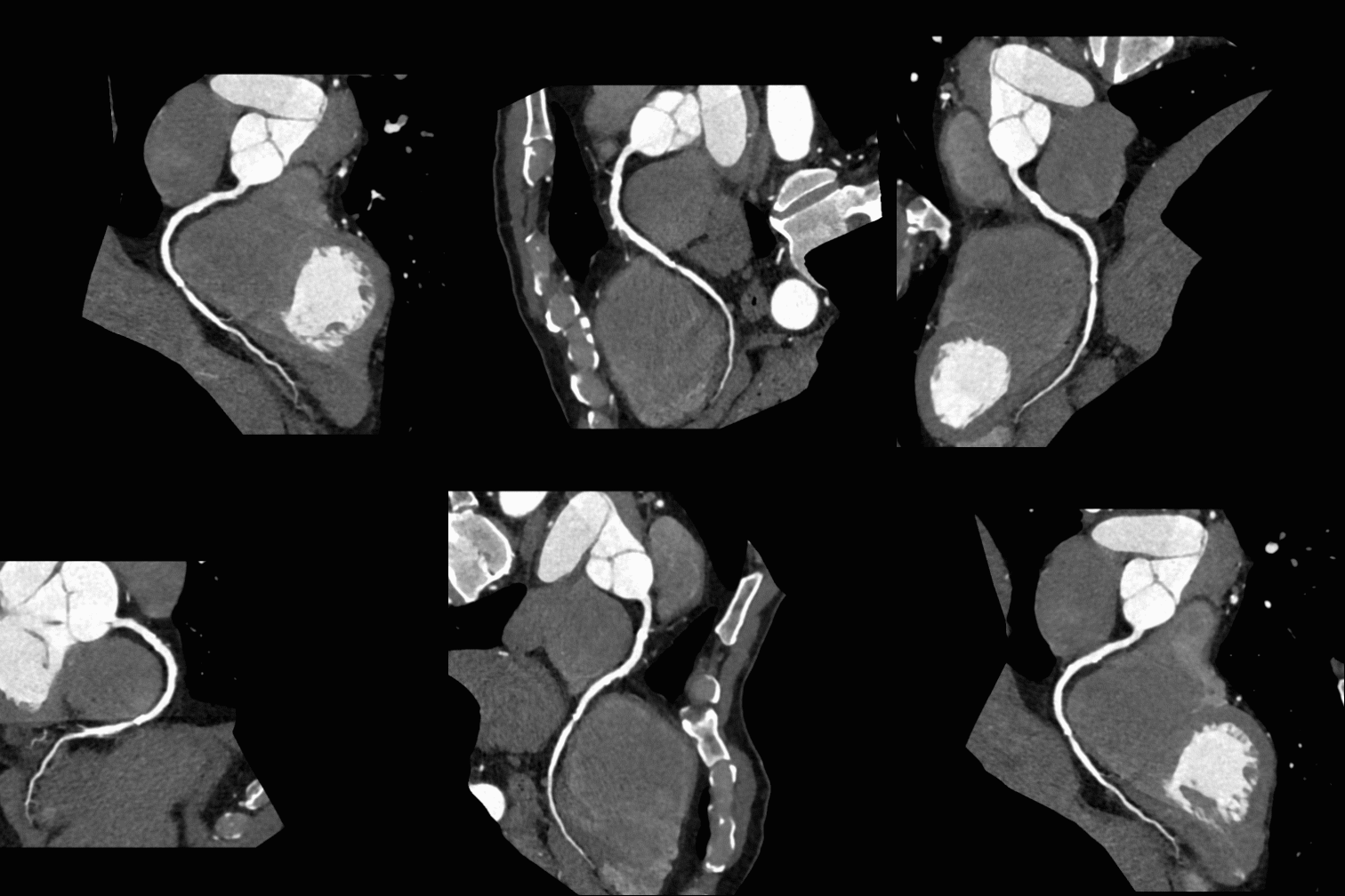
While raw medical scan data is valuable, it often comes with constraints such as being limited to three standard views (sagittal, coronal, axial) and is sometimes limited in scope or range due to scanner capabilities or the nature of the scanning modality. 3D imaging addresses these issues through various image processing techniques. These methods enable the creation of custom, non-standard views, allow for the fusion of different data types, and enable the stitching of aligned scans, among other enhancements. These techniques broaden the scope and utility of medical imaging, overcoming inherent limitations of the original scans.
Batching
Curved Planar Reformatting
Image Fusion
Stitching

Measurements & Surgical Planning

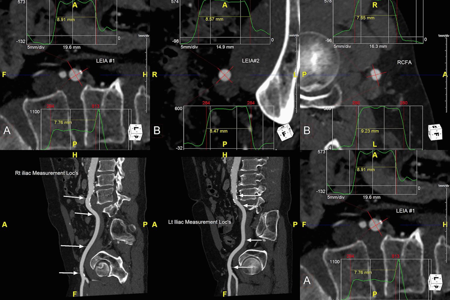
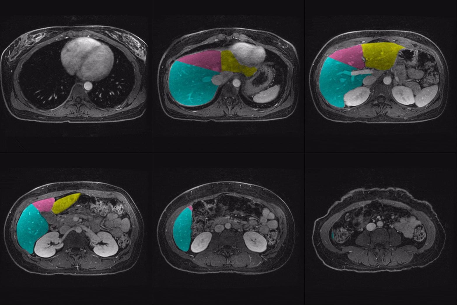
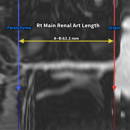
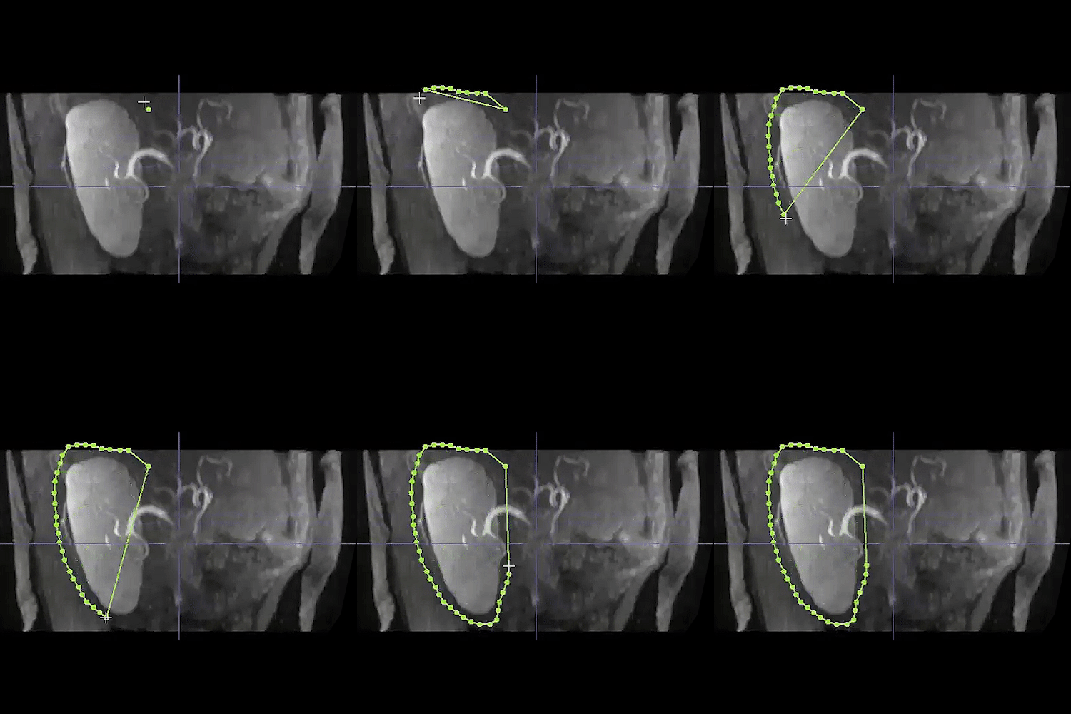
Representations of a patient’s anatomy are important for surgical planning, and are greatly enhanced by precise measurements from scan data. This level of precision is particularly beneficial for understanding the complexities of individual anatomies. By utilizing measurements, surgeons can plan their procedures with higher accuracy, foresee potential challenges, and strategize the most effective approach for surgical intervention. The ability to generate patient-specific 3D imaging aids surgeons in planning complex surgeries, significantly reducing the risk of complications during the actual procedures and improving surgical outcomes and patient safety.
3D Imaging - adrenal hypertension

Common Measurements
Kidney Volumes
Vascular Distance Measurements
Liver Volumes

Advanced 3D Imaging

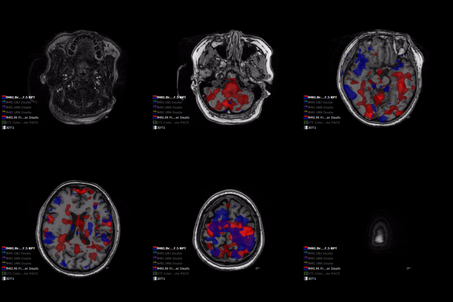
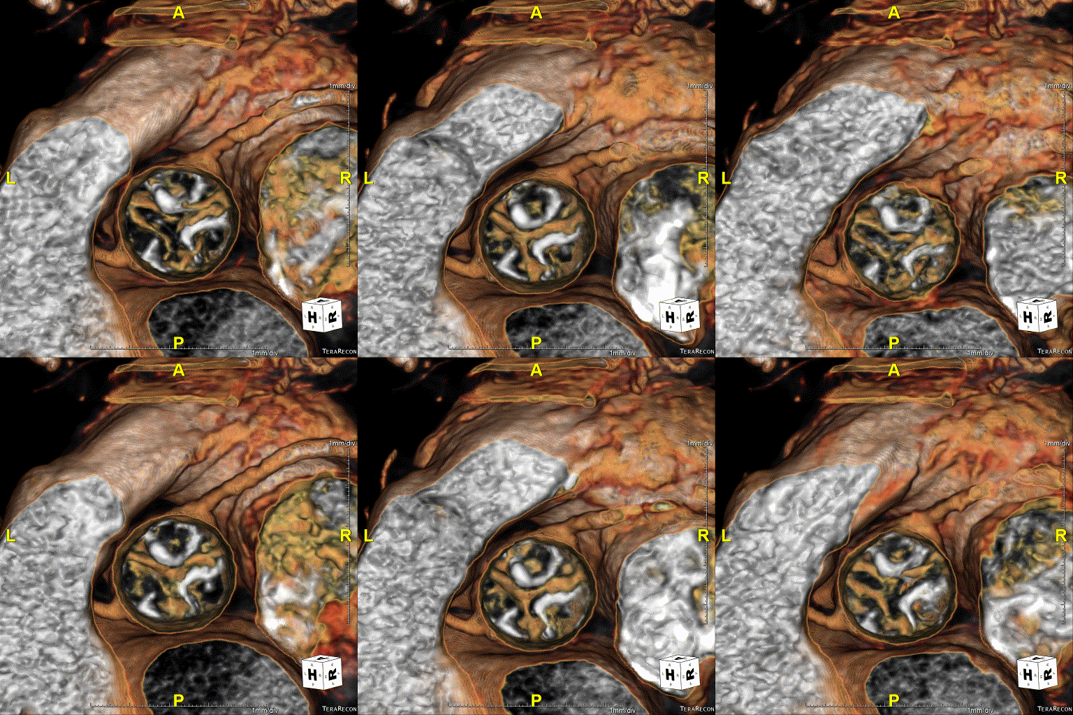
Many advanced techniques go beyond the 3D imaging techniques described above, each offering unique advantages. Among these, 4D imaging introduces the dimension of time to 3D images, allowing for the observation of anatomical motion and changes over time. Functional MRI (fMRI) generates 3D structural images of the brain, supplemented with functional data to map brain activity. 3D colonoscopy, also known as virtual colonoscopy, creates a 3D model of the colon for detecting polyps and cancers, serving as a less invasive alternative to conventional colonoscopy. These examples are just a fraction of the advanced 3D imaging techniques available, with many others being used and developed in the medical field to enhance diagnostic and treatment capabilities. 
4D Visualization
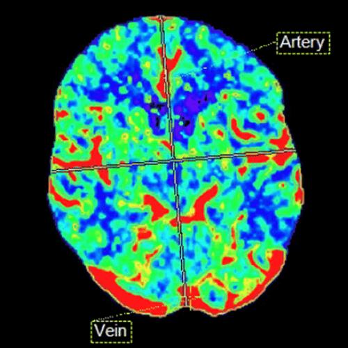
AI Stroke Analysis
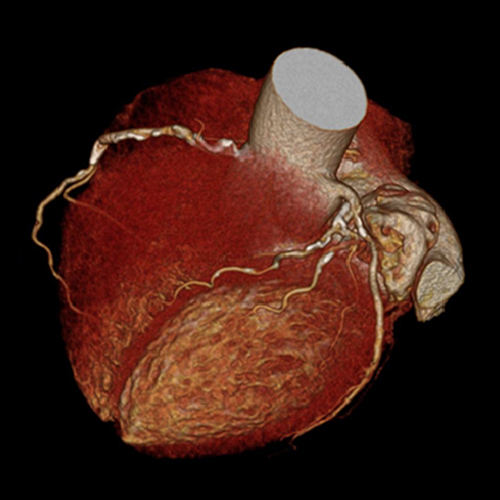
Calcium Score
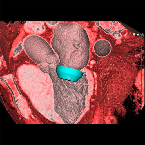
Device Simulation

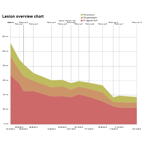
Longitudinal Analysis

Longitudinal analysis enhances 3D imaging by providing information about changes in the body over time. This technique allows for precise monitoring of the progress of a medical condition, such as tumor growth or aortic aneurysm. By comparing 3D images of the same anatomical part, taken at different intervals, doctors can accurately assess the effectiveness of treatments, like the impact of chemotherapy on a tumor. This temporal visualization aids doctors in making informed decisions about starting, changing, or continuing treatment strategies. 
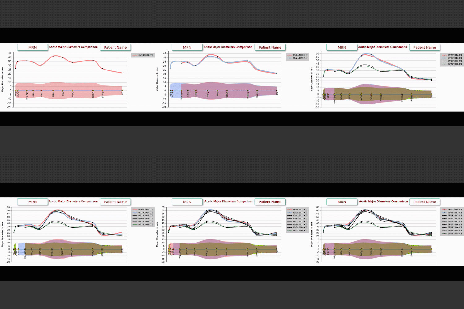
Aortic Surveillance
Tumor Response Assessment Criteria
Tumor Quantification
In conclusion, the value of 3D imaging in healthcare is reflected in its adaptability to various medical needs. From enhancing diagnostic accuracy to providing surgical guidance, 3D imaging technologies provide a unique benefit in medical practice. It is with 3D imaging that complex medical data can be unlocked, making it more personalized and effective. As the healthcare community continues to embrace and refine these technologies, sharing insights and advancements becomes crucial. By sharing knowledge and experiences healthcare professionals worldwide can participate in this transformative journey, collectively advancing the field and ultimately benefiting patients across the globe. The future of 3D imaging in healthcare is not just about the technology itself, but about fostering a collaborative environment where information and resources are shared, paving the way for innovative solutions and improved health outcomes.

(1) Learn more about CT Imaging quality here.
(2) Learn more about MR Imaging quality here.

Categories

  • Head & Neck
  • Chest
  • Abdomen & Pelvis
  • Upper Extremity
  • Lower Extremity
  • Case of the Month
  • Techniques

3DQ Lab – Grant Building

300 Pasteur Drive
Stanford, CA 94305

(650) 725-8432

Directions to Grant

3DQ Lab – Clark Building

318 Campus Drive
Stanford, CA 94305

(650) 725-6862

Directions to Clark

Learn More About the Lab

  • About Us
  • Meet the Team
Search Search

Copyright © Stanford University

© Copyright - 3D and Quantitative Imaging Laboratory - Enfold Theme by Kriesi
  • Link to LinkedIn
  • Link to Instagram
Link to: May Thurner Syndrome Link to: May Thurner Syndrome May Thurner Syndrome Link to: 3D Printing: Pegs Link to: 3D Printing: Pegs 3D Printing: Pegs
Scroll to top Scroll to top Scroll to top