Innovation Projects

The 3DQ Lab’s work extends beyond routine case processing to support its broader mission in education, research, and patient care. Alongside clinical production, a wide range of projects focus on developing new approaches to imaging analysis, advancing 3D modeling and printing, evaluating and integrating AI tools, and improving how imaging data is visualized, measured, and communicated.

Many of these efforts begin with a specific need, whether refining a workflow, supporting a clinical service, enabling a research study, or exploring a new technique. While they are not part of standard case output, they often build directly on clinical experience and are designed to improve consistency, expand capabilities, and support more informed decision making across teams.

This page highlights those projects, from patient-specific 3D printing and quantitative imaging methods to AI development, protocol design, and collaborative research. Together, they reflect how innovation within the lab supports both day to day clinical work and ongoing academic efforts.

Patient-specific 3D printed prostate slicing guides helped standardize how specimens were sectioned to better match MRI, supporting automated alignment with pathology, improving dataset quality for AI, and enabling more accurate comparison to refine lesion grading.

With fewer mediastinoscopy cases for resident training, a patient-specific 3D printed mediastinum model was created for practice. Residents reported increased confidence, demonstrating how tailored 3D printing offers repeatable procedural training when clinical volumes are limited.

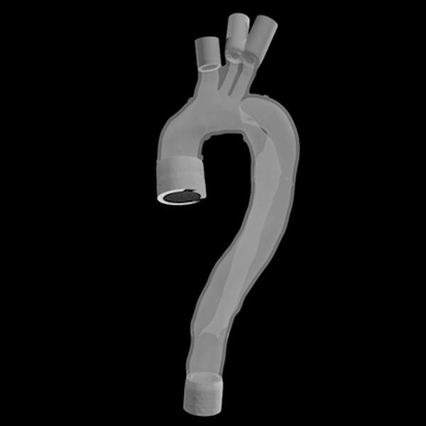
Patient-specific aortic dissection models were 3D printed to support 4D-flow MRI and fluid-structure interaction research. The 3DQ Lab contributed design modifications, multi-material printing workflows, and durable coatings to better replicate physiologic aortic behavior.

Patient-specific 3D printed models were created from CT angiography to support microsurgical reconstruction. Their use reduced flap harvest time and improved confidence in breast reconstruction, burn reconstruction, and vascularized lymph node transfer procedures.

Patient-specific heart models were used to simulate how septation repair would affect circulation. The 3DQ Lab created cardiac segmentations and designed and 3D printed four molds used to cast silicone hearts for experimental computational flow studies.

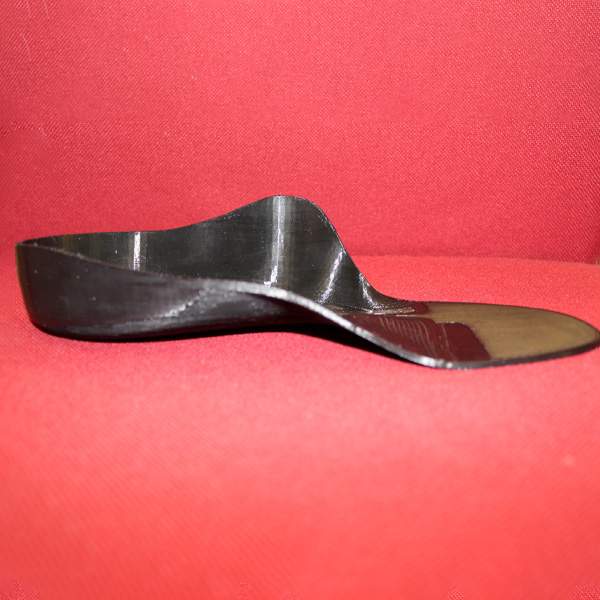
In collaboration with Orthopaedic Surgery, the 3DQ Lab designed a patient-specific orthotic for a complex reconstruction case that traditional orthotic providers were unable to support. Initially using a 3D scan for reference the design has been iterated upon many times.

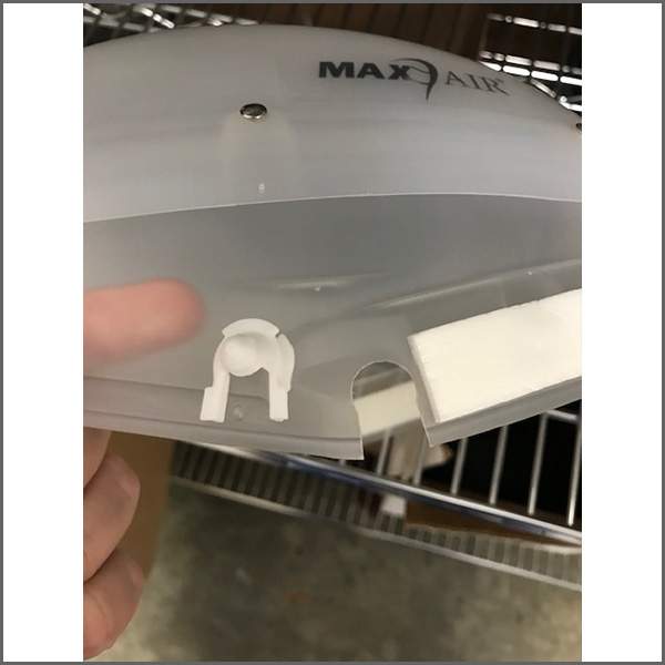
During the COVID-19 pandemic, the 3DQ Lab reverse engineered and 3D printed replacement parts for CAPR systems after OEM components became difficult to obtain. Hundreds of parts were produced and delivered to help maintain respiratory protection supplies across Stanford Health Care.

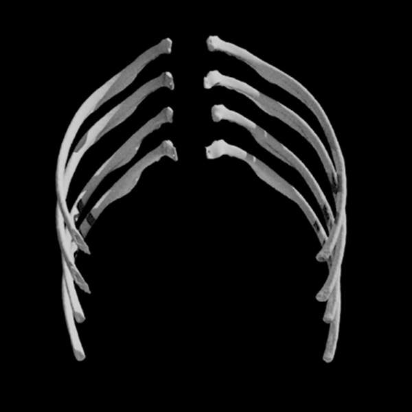
For a complex Neurosurgery case, the 3DQ Lab created full-scale spine and rib models to support development of a custom stabilization device. Anatomically accurate and modified rib versions were printed to evaluate the patient’s anatomy and potential attachment strategies.

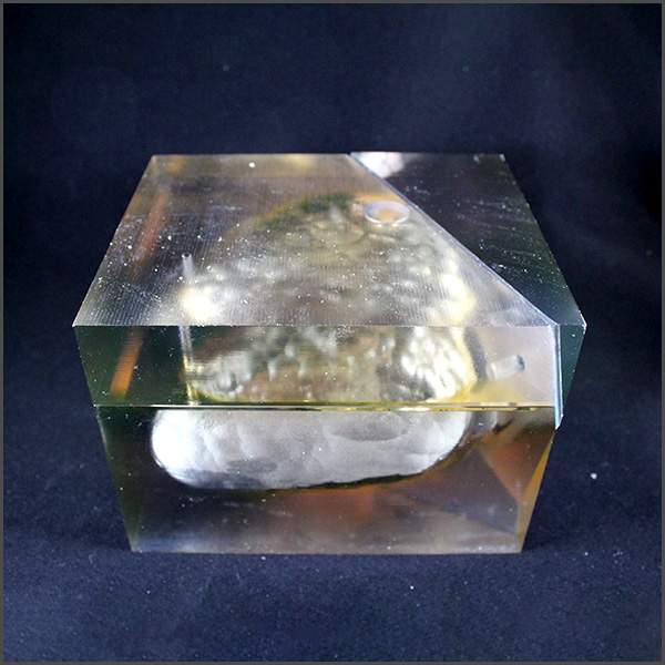
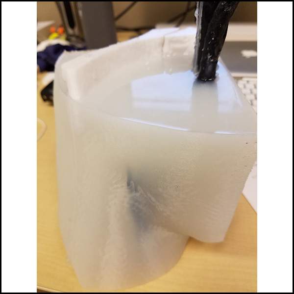
The Stanford 3DQ Lab collaborated with Interventional Radiology to create reusable vascular access phantoms. The process involved segmenting vasculature, 3D printing, and ballistic gel casting to create an ultrasound-compatible trainer for new residents and physician trainees.

The Stanford 3DQ Lab collaborated with Cardiothoracic Surgery to develop a multi-material chest wall model for Nuss procedure teaching. The model combined rigid bone anatomy with flexible costal cartilage to support patient education and fellow training.

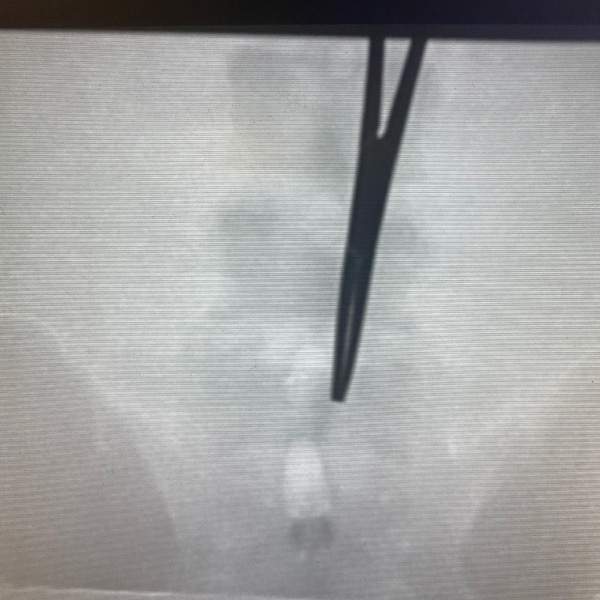
A reusable fluoroscopy-compatible lumbar puncture phantom was developed in collaboration with Stanford General Surgery as an alternative to commercial training models. The project evaluated materials, infill strategies, and fabrication methods for image-guided procedural education.

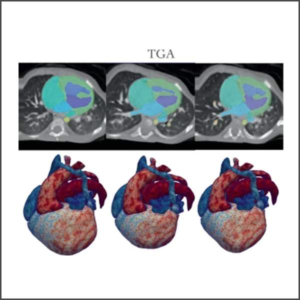
A deep learning model was developed to generate synthetic 3D heart anatomies for rare congenital heart diseases. These virtual datasets can support segmentation training, computational simulations, and future research when real patient cases are too limited for robust AI development.

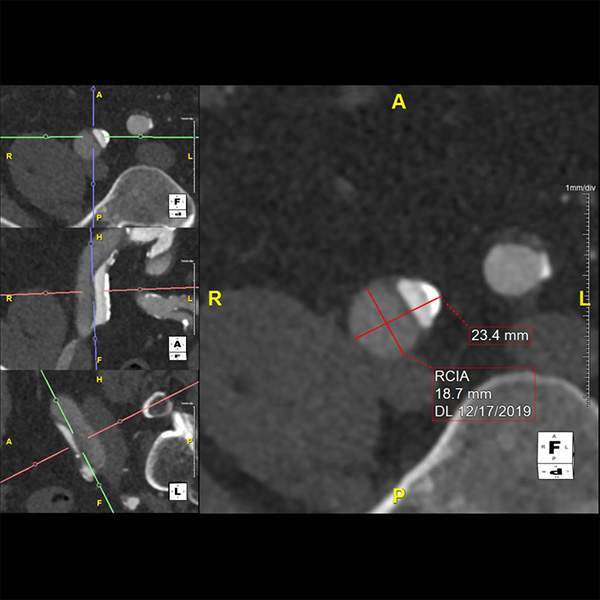
The 3DQ Lab developed a workflow to extract measurements directly from FDA-cleared 3D imaging software scene files, reducing manual entry while enabling structured clinical data to be reused for retrospective analysis, machine learning, and other research applications.

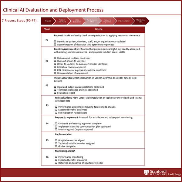
A structured program was developed to support the evaluation, deployment, and ongoing management of artificial intelligence tools in medical imaging. Standardized workflows, lifecycle tracking, and dedicated support help guide algorithms from initial testing through clinical implementation.

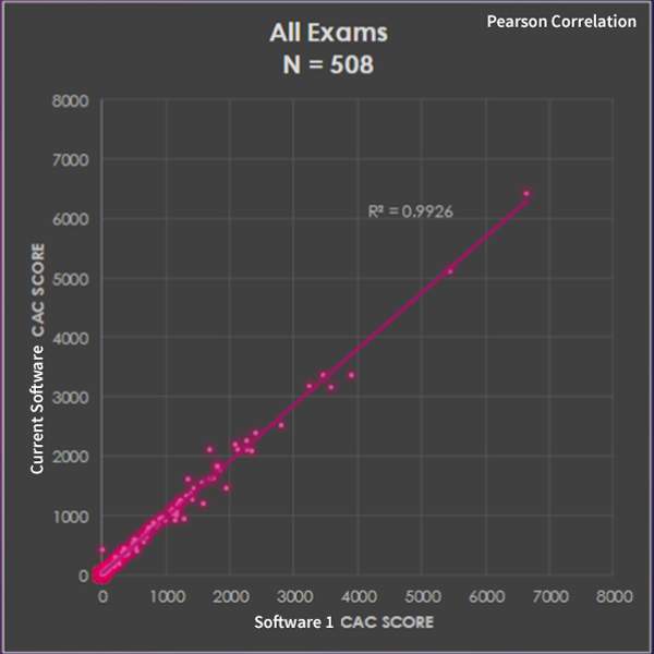
Automated calcium scoring software was evaluated and compared against current manual workflows. Strong agreement between the results supported integrating the automated solution into 3DQ Lab processes to improve turn-around time and shift technologists towards other specialized needs.

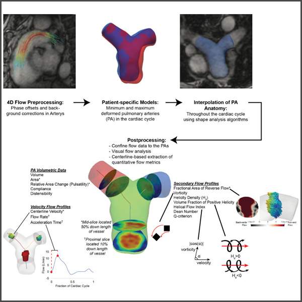
A semi-automated pipeline was developed to analyze 4D flow MRI of the pulmonary arteries before and after pulmonary endarterectomy, linking changes in vessel stiffness and blood flow patterns with improvements in pulmonary pressures and right ventricular recovery.

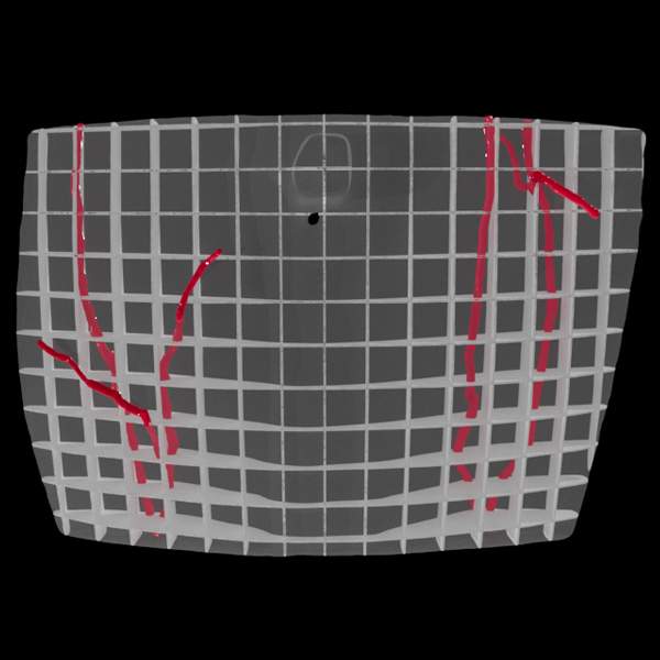
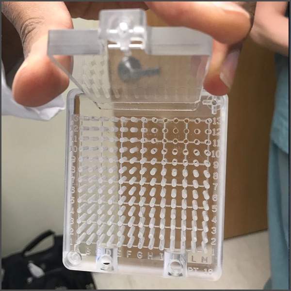
Developed with the Department of Urology, this reusable needle guidance grid helps position and stabilize needles during transperineal prostate biopsy and cryoablation. The 3DQ Lab led the design refinement, 3D prints the grid and attachments, and has supported more than six years of clinical use.

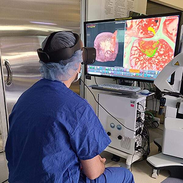
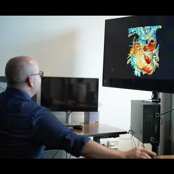
The 3DQ Lab provided in-house support for Neurosurgery’s Surgical Theater platform, creating patient-specific 3D models and custom workflow tools to support surgical planning, case coordination, and virtual reality review in both clinic and operating room environments.
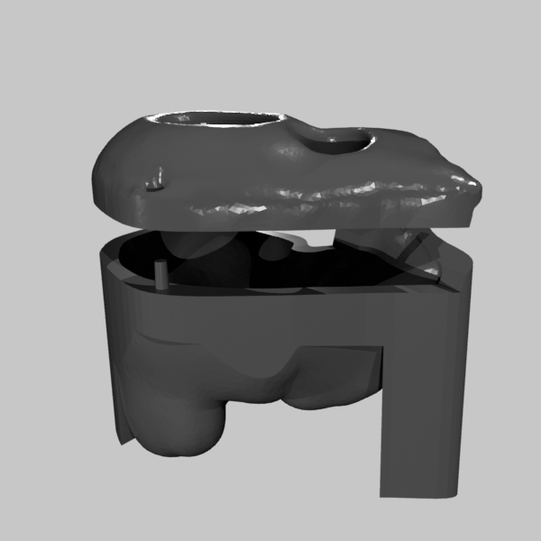
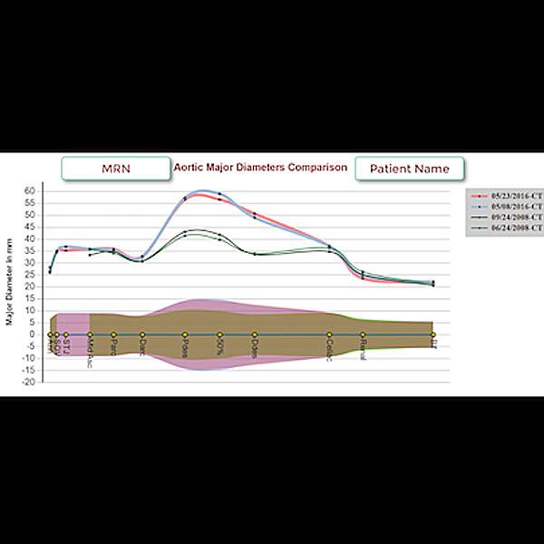
The 3DQ Lab developed an aortic surveillance workflow that standardized measurements, automated graphing, and streamlined communication with Cardiothoracic Surgery, providing rapid access to imaging data for long-term monitoring and surgical decision-making.

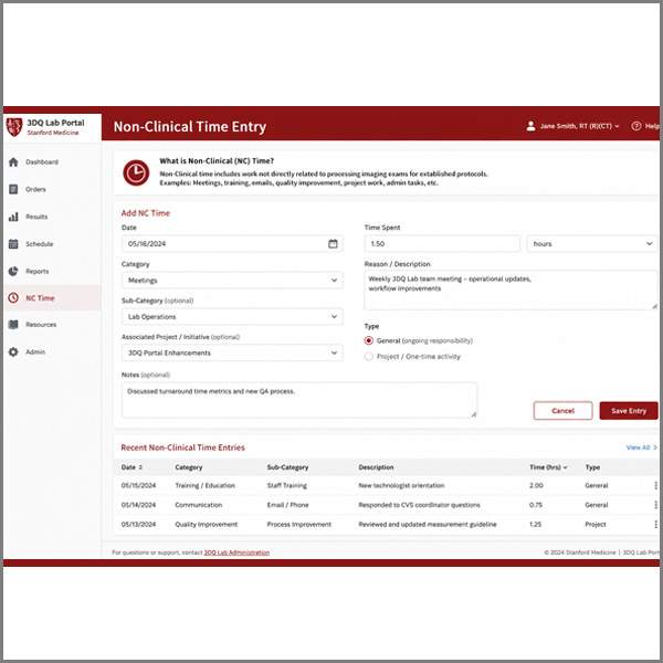
The 3DQ Lab developed a system to track NC Time (Non-Clinical Time), capturing work such as meetings, training, and communication that is not reflected in exam counts. This created a more accurate approach to measuring staff productivity and overall contributions.
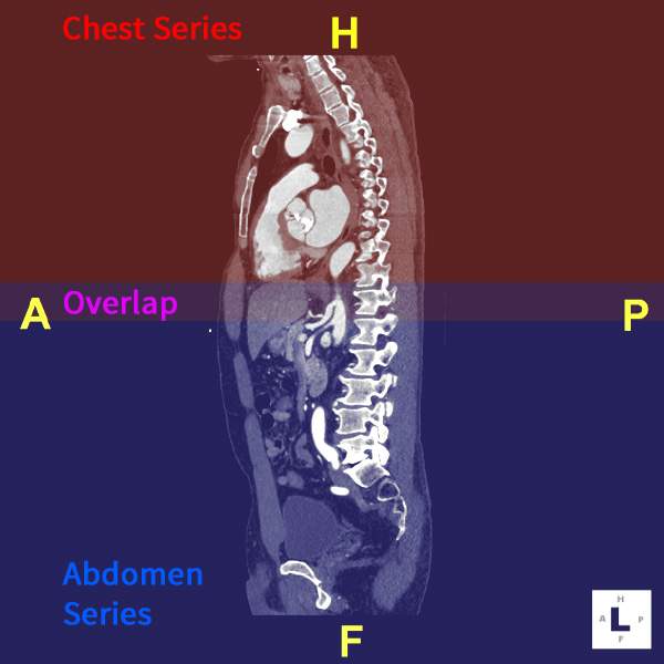
The 3DQ Lab explored an automated workflow to stitch multipart CT angiography studies and batch process them for PACS delivery. Although the project was not ultimately adopted, it demonstrated the lab’s efforts to reduce manual processing and improve imaging accessibility.

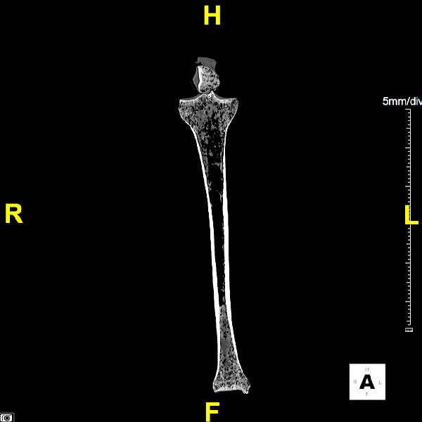
A standardized CT protocol for orthopedic allografts was established to support patient-specific surgical cutting guide creation for musculoskeletal tumor reconstruction. The workflow integrates donor bone imaging into existing PACS and surgical planning systems.

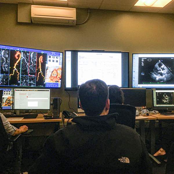
The Stanford 3DQ Lab supports TAVR planning meetings by providing live imaging and to the medical team, helping clinicians collaboratively evaluate anatomy, vascular access, valve sizing, and procedural considerations before intervention.

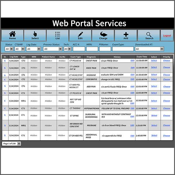
Protocol documentation resources, imaging software workflows, and web portal elements are maintained by the Stanford 3DQ Lab to help standardize imaging outputs across our many technologists while adapting to evolving clinician preferences and operational needs.

Aorta surveillance reports can be converted into DICOM format for PACS storage, creating a pathway to reduce manual report distribution while improving access to longitudinal measurement data within existing clinical imaging systems.

The Visiting Fellowship Program provides individualized training in clinical image post-processing, quantitative imaging, laboratory operations, and medical 3D printing, helping imaging professionals gain practical experience with advanced visualization and clinical workflows.

Dedicated 3DQ Lab staffing within Stanford Cardiothoracic Surgery provides immediate imaging support during patient consultations. Direct access to post-processing expertise helps answer imaging questions in real time and improves communication between imaging and clinical teams.

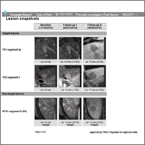
Tumor-directed immunotherapy requires consistent measurements tracked over time for accurate response assessment. Stanford Radiology partnered with the Stanford 3DQ Lab to create TRAC, which provides standardized longitudinal measurements for response assessment and planning.

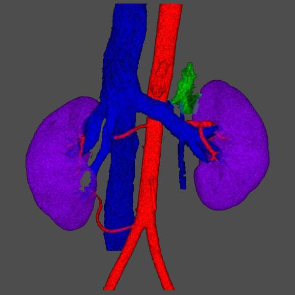
A new augmented reality rendering method was evaluated for interactive 3D visualization of kidney donor vascular anatomy on AR headsets. Surgeons reported higher confidence assessing renal arteries, while the 3DQ Lab supplied the segmented patient-specific 3D meshes used for visualization.

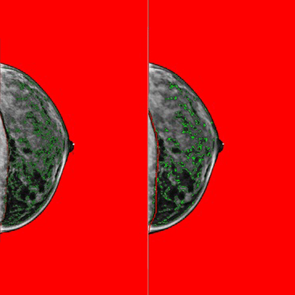
Processed digital mammograms were evaluated to confirm reliable breast density measurement using Cumulus. The 3DQ Lab supported large-scale analysis by processing over 30,000 cases and building data workflows, enabling practical risk assessment from routine clinical imaging.

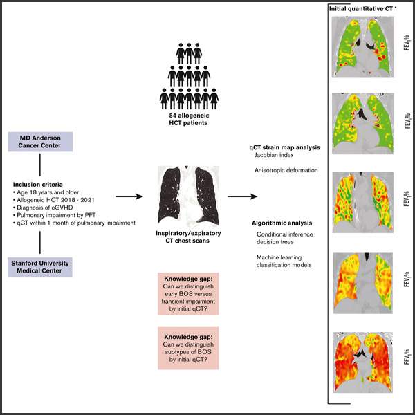
Quantitative CT was used to detect bronchiolitis obliterans syndrome earlier than standard methods. 3DQ Lab–generated IMBIO 4-color outputs highlighted regional lung changes, supporting diagnosis, disease classification, and consistent analysis in transplant patients.

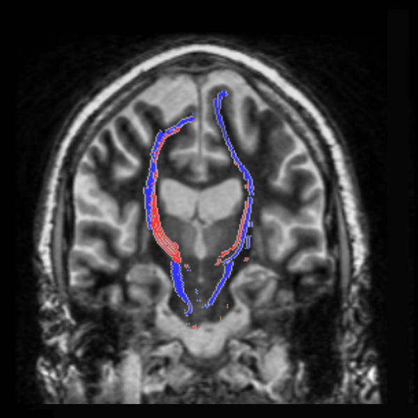
Focused ultrasound treatment for essential tremor requires consistent localization of the VIM region and nearby motor pathways. Stanford Neuroradiology partnered with the 3DQ Lab to create a DTI tractography workflow for DRTT mapping and standardized outputs for procedure planning.

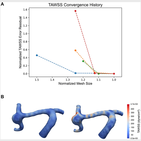
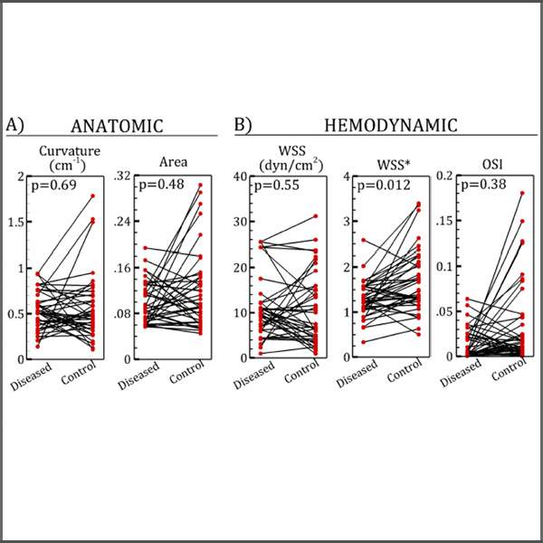
Cerebral aneurysm growth patterns were studied to identify blood flow factors linked to progression. Using computational simulations of matched cases, the study compared growing and stable aneurysms, with the 3DQ Lab providing segmented vascular models for analysis.

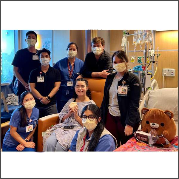
A patient-specific 3D printed heart model was created as a physical keepsake for a pediatric transplant patient at Lucile Packard Children’s Hospital, providing a tangible reminder of her journey through heart failure, transplantation, recovery, and healing.

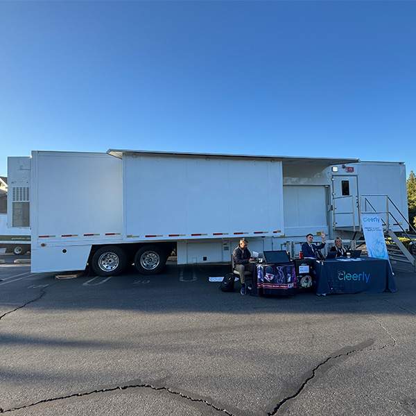
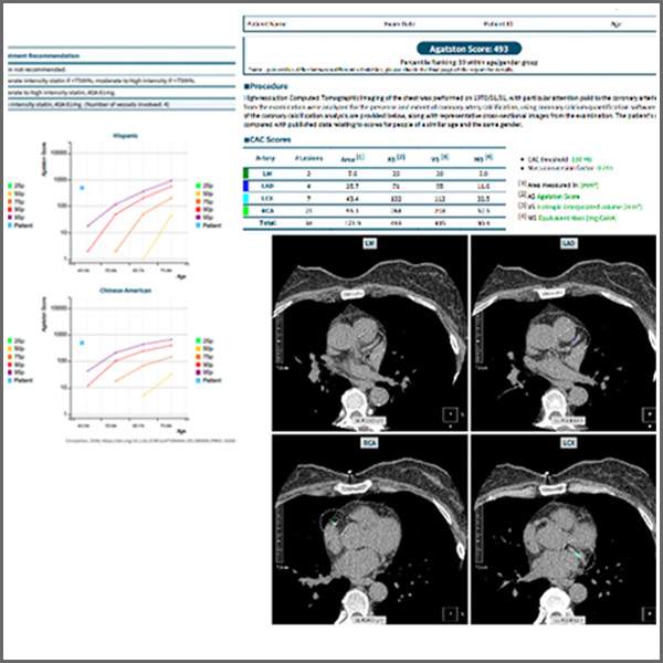
The 3DQ Lab supported an off-site coronary calcium scoring event by coordinating patient flow, transferring images from the mobile CT scanner to PACS, and performing the calcium scoring analysis. Customized routing within the imaging portal helped separate event studies from routine clinical imaging.

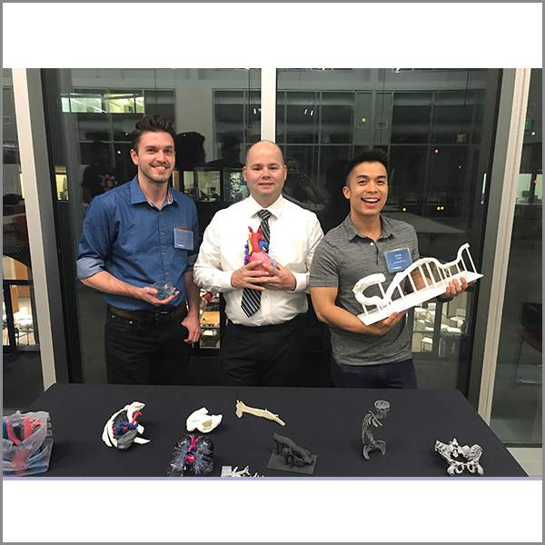
Members of the Stanford 3DQ Lab have cumulatively delivered more than 80 presentations, workshops, and educational sessions at national meetings, institutional programs, and student events, sharing practical approaches to advanced imaging, 3D printing, and clinical workflow development with a wide range of audiences.

Patient-specific 3D liver models were evaluated to address size mismatch, a major cause of organ refusal in pediatric transplant. Silicone cast models from printed molds enabled accurate volume assessment, improving donor selection, planning, and surgical outcomes.

The 3DQ Lab generated approximately 75 patient-specific models used for computational analysis of bypass graft failure in collaboration with the Marsden Lab. Lower wall shear stress was associated with regions that developed stenosis, supporting identification of high-risk grafts.

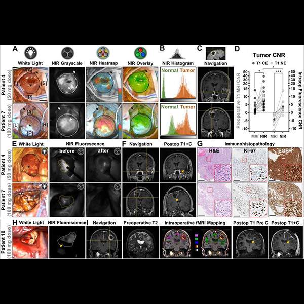
Fluorescence-guided glioma surgery used near-infrared imaging and MRI correlation to improve visualization of tumor tissue during resection. The 3DQ Lab supported imaging overlays and functional MRI workflows used to correlate fluorescence signal with tumor location and surrounding anatomy.

The 3DQ Lab processed more than 1,000 coronary artery calcium scoring studies for Stanford’s participation in Project Baseline, a landmark longitudinal study designed to better define health and create a research dataset that continues to support cardiovascular research.

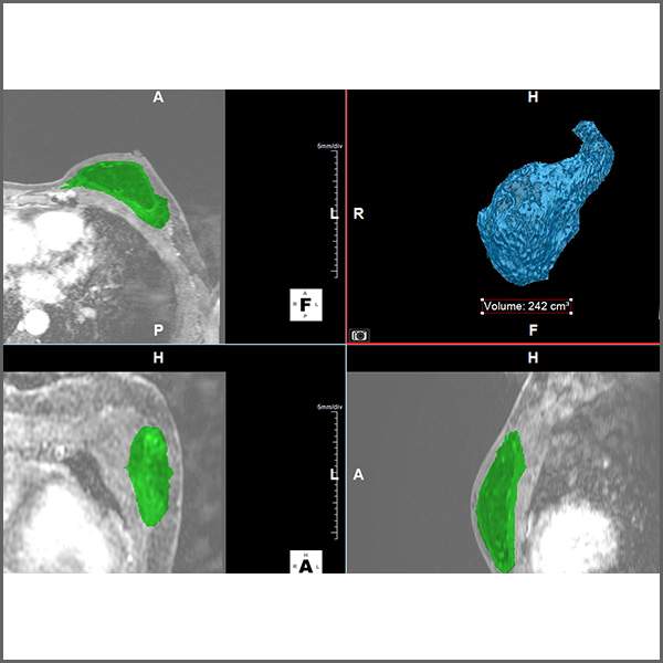
Working with the Division of Plastic and Reconstructive Surgery, the 3DQ Lab developed a method to measure breast volume from CT imaging, providing quantitative data that supported publication and broader adoption of Stanford’s novel omental flap breast reconstruction technique.